Editorial Board Comments:

Samir Pancholy:
I have seen for so far, all return on physical exam, no complaints from the patient, all evaluated by vascular surgery as well, and left alone. Besides prominent veins that I noticed only after I knew the diagnosis, there was no other consequence. All of them are in their mid to late 80s. No intervention needed. Hope this helps.

Sunil Rao:
I would jus observe. These usually dont end up causing problems.

Kimberly A. Skelding:
Agree would observe.

Rajiv Gulati:
Our single-center data: AV fistulae N = 3/10540 TR caths. All were treated surgically and did well (apparently surgery is q straightforward). That said, a period of observation seems very reasonable if asymptomatic. Do give us follow-up!

Ian C. Gilchrist:
Not life-threatening and usually small, would leave alone. If patient bothered, the surgical fix is easy (although is local surgery). May be able to compress some closed especially if not on anticoagulants/ antiplatelets but have no experience here.

Christopher T. Pyne:
I think we have had a couple (not mine of course!!) – they got operated on and did well – all were complaining of pain in their wrists…

Yves Louvard:
Dear friends, I never had one (around 30.000). So as others I can say it is extremely rare. One of my associate had one which was small and untouched Jean Fajadet In a workshop in Massy reported a long time ago a case he sent to a surgeon, no more detail… and no recommendation.

Alejandro Goldsmit:
Dear fiend, in more than 10 years, Never.!!!!, no recommendation. im sorry.

David E. Kandzari:
I would suggest observation as well, although I would try every measure before surgery if symptomatic.

John Coppola:
I had one just watched it my partner had one fixed by vascular under local same day procedure. I would watch.

Mitchell Krucoff:
If it became symptomatic seems like US guided compression would be an interesting option, as the vascular structure is so easy to visualize, but I have never done.

Kintur Sanghvi:
I had a patient recently mostly asymptomatic but concerned, applied USG guided Radar band with high compression pressure for 4 hours. It was successful. In past about a year ago on a different patient similar attempt was unsuccessful. That patient ended up getting a small surgery while undergoing CABG.

Mauricio G. Cohen:
I only had one case, diagnosed in the office by the presence of a thrill and a bruit, and confirmed by ultrasound. Completely asymptomatic, although the patient was anxious. We did nothing. Just observation. I dont feel strongly about watchful wait versus surgical intervention, but if asymptomatic and not functionally relevant, first do no harm. Good luck with your case.

Ronald Caputo:
I have never seen one – probably because I never look for them and they have no symptoms.
There was a patient across town that they called me about – the patient could feel the flow through the fistula. I suggested compression and it luckily worked. Hope all of you are well.

Tejan Patel:
I have one patient of mine has same thing. Pt. is asymptomatic. No need for treatment in my opinion. It was funny one of the vascular surgeon who show the pt. Mentioned if we can figure out to creat asymptomatic AV fistula like this and may be it will mature enough in future for Dialysis!!!!

Tejas & Sanjay:
Radial arteriovenous fistula:

  • If fistula is very small, it can be managed conservatively.
  • If it is moderate to large sized, it should be managed surgically.

David Hildick-Smith:
Thanks to everyone so far for these extremely helpful responses from the Worlds Transradial Experts!


Editorial Board Comments:

Ian C. Gilchrist:
Interesting. Surprised there is no high-output failure. Never seen anything like this but must be some sort of syndrome.

Sunil Rao:
There was a similar case at Duke recently. That patient had an RCA from the PA but survived to age 47 because of a gigantic 5 mm diameter collateral from a huge apical LAD. Eventually she developed right heart failure and required reimplantation of the RCA.

Olivier Bertrand:
We had quite a similar sized fistula going from Cx and RCA to RA tumor. We did embolize from both Cx and RCA prior surgery. There is also a similar case published recently in JACC interv.


Kintur Sanghvi:
Coronary fistula to tumors or cancer are more common than such large congenital fistula which most commonly connect to the main pulmonary trunk  or left pulmonary artery trunk. Most common reason for such large fistula is pulmonary displasia at an early age in childhood or in-utero. I presented one such case at the Angio session at TRICO 2012. That patient had Coarctation of AO and coronary fistula to PA. I have attached the AVIs here. In fact I have taken care of 3 such patients (as Deborah had a large and robust pediatric cardiology department in the past and patients still follows with us). Trying to coil embolize such large fistula is not feasible and patient continues to grow new fistulas to PA from other arterial system. One such example is published by my old partner. He tried to treat with coils and put over 100s of coils in different fistulas in that patient and patient still keeps coming back with new fistulas. That case was published: www.cathlabdigest.com/articles/Chest-Pain-Secondary-Recurrent-Coronary-Pulmonary-Artery-Fistula. The surgical correction is relatively an easy operation by ligating the fistulas and patch repair of the main pulmonary trunks. The patient I am sending the AVIs for was treated that way in 2011 and have done well. But as I searched literature more for the last two patients I have seen and I am treating both of them conservatively. It is not necessary to treat these patients as they do not get ischemia of myocardium if you study with nuclear exercise stress test. Despite these large fistula the coronary physiology is not altered!!!Here is an editorial from Texas Heart Journal that I have used as a reference. https://www.ncbi.nlm.nih.gov/pmc/articles/PMC101097/#r2-1

Editorial Board Comments:

Rajiv Gulati:
Dear Mitch – yes. We frequently do such venoplasty to assist our EP colleagues with complex pacemaker re-dos. Typically a 5 or 6 mm coronary/peripheral balloon will suffice although you can go larger. Two complications in our early experience a few years ago – from dilation after wire exit with a stiff glide. Both times wire passage had been tough.

Samir Pancholy:
Dear Mitch: Yes, venoplasty with low pressure inflation can be done without trouble especially in a fibrosed vein with a foreign body in it. I have done in several times in the subclavian (even in the non-compressible zone) to advance large ICD leads with previous pacing leads in place. We also use balloon dilatation in coronary veins for LV lead placement without difficulty. I have never done it for placing a swan although. The key is to keep pressure <6 atm. Usually recoils by the end of the case.

Ian Gilchrist:
Another option is to go smaller, use a 4-F Balloon wedge.

John Coppola:
Agree with Sam have done for our ep people in several renal failure patient who had fibrosis subclavian from prior dialysis catheters. Use 5 to 6 mm balloon low pressure the pass lead by end of case recoil.

Ronald Caputo:
I assume you are going from the left arm? you can definitely dilate this safely but I would use a very low profile balloon 5 mm diameter (5×40 or 5×80 mustang)I have dilated and stented the SVC with really large balloons and at high pressure but only in the setting of SVC syndrome  – often post radiation so similar situation re fibrosis.  I would not dilate at super high pressure due to concern for rupture The other option would be to try the other arm (right) and cruise past the PIC

Mitchell Krucoff:
Is everyone comfortable with RONs suggestion to go in the PIC line arm as an option?

Ian Gilchrist:
I have done that with problems. Aim for different vein.

Samir Pancholy:
Agree. Should workout fine. There is plenty of room proximally to accommodate both catheters.

Tejan Patel:
No, As we all know all road ultimately leads to Rome (SVC) so left or right does not matter.  Long term solution is remove fibrous cap and Laser Athrectomy is best solution. Best solution in my opinion pass some 5-6F sheath/Guide catheter through the CTO of SVC and flot catheter through it. I have done it once so far. Again Balloon Assisted Technique works even in these situation!!!

Mauricio G. Cohen:
I have been passing balloon tipped catheters through the same side as a PICC line without any difficulties. Our access is usually distal to the PICC insertion and there is always room for the Swan and the PICC in the basilic/ brachial vein.

Tejas & Sanjay:
We should avoid right subclavian route. Dilation of subclavian vein may dislodge lead. Femoral route is preferable.

Editorial Board Comments:

Jennifer Ann Tremmel:
It is a fairly common problem and a JL3 is what you need. I consider it a “must-have” in any radial lab.

Yves Louvard:
I agree Jennifer but not always available. Other option an AL. Very good year 2016 to all of you.

Sunil Rao:
JL3.0 (usually available only as a guide catheter) or a JCL catheter by Cordis will work. For PCI, you can use a JL3.0 guide but now Cordis offers an XB2.5 guide catheter as well.

Olivier Bertrand:
My routine is JL 3.5, If that does not work plan B is either a guiding XB 3.0 or 3.5 (5Fr) depending of aorta size or Amplatz.

Samir Pancholy:
XB 3.5 works for me nearly always. If not, we use XB3

Sunil Rao:
Samir and Olivier, for the scenario described below where there is a narrow aorta and a short distance from the cusp to the left main (which is also posterior), I have never been able to get an XB3.5 to fit. It is just too large. What is your secret to getting these to work? Also, while we are on the subject, what is the communitys catheter of choice for engaging the RCA when there is a lusoria? I have often struggled and used a JR with the 0.035 wire in it to prevent kinking during the aggressive torquing. Nowadays I pretty much bail to the left radial when I encounter a lusoria.

Tejan Patel:
We have JL 3.0 by Cordis diagnostic catheter for more than 5 years available in Cath lab. I think it is special order and it will work in this situations. I agree. If nothing works go to basic try anything that works before puncturing other site!! Do not forget LIMA catheter.

Ian Gilchrist:
When the JL3.5 does not work, I proceed with the JR4 to see the RCA and then turn the catheter around and see if it reaches the LCA. Works at times and avoids taking out the whole inventory. Does not help for PCI, but how often do young women need PCI? Working too hard. Use the left radial.

Olivier Bertrand:
We have asked Cordis to produce XB 2.5 (Not sure they still do it since acquired…)…Not used it for a couple of years. For lusoria always pain in the from the right, Used Amplatz and extra stiff wire (usually multiple exchanges), Left is probably the most straightforward way too.

Tejan Patel:
Sunil, I try JL 3.5 for left for arteria lusoria and some time you can shape JL to seat in RCA with 0.35 stiff end of it does not work than JR 4 or even 3D RC works. Of course first I would have tried with Tiger just to find lusoria! Sometime even Tiger works with some difficulty. Though better for RCA rather than LM.

Samir Pancholy:
Good questions. I usually initially, enter LV with EBU and pull back into LM, works better. If fails, I use the stiff end of 0.035″ wire to modulate the EBU curve and engage. Support of EBU is much better than JL. For diagnostic only, JL has worked. Tiger would work too, for diagnostic cases. For right coronary, my go to catheter is always MAC, in this case with the wire. That lets me shape it from JR to Hockey to MP etc. just by advancing or withdrawing the wire. Once in the backup is great. I tell the senior fellows that a “pseudolusoria” with short ascending and small root is their graduation case.

Sunil Rao:
Cordis does still produce an XB 2.5. Can be valuable in specific cases.

John Coppola:
You can get a special order diagnostic JL3.0 or since it is impossible to order a 8 dollar diagnostic catheter in a city hospital I use an 85 dollar EBU 3.0 which works well.

Rajiv Gulati:
Great tips and tricks. An up vote to Ians suggestion of JR4 for LCA in short narrow aortas. Has worked well for me, inadvertently. Re right from lusoria a provokes operator spasm. I recollect some success with MP and AL3 but more often I have switched access.

Kintur Sanghvi:
Happy New Year Friends, JL 3.0 diagnostic and JL 3.0 (guide Launcher) both available for 5 years since I have been at Deborah. BSC, and Cordis have JL 3.0 as a choice in their catalog of diagnostic catheter. Similarly when a RCA is originating from the left cusp, JL 3.0 works very good.

Ronald Caputo:
I do not run into this often and use the sones technique for a majority of my diagnostic but I think that pre-formed JL-3, AR-2 or AL 0.75 are worth a shot. Cannot use a JR with lusoria. The best bet is an Amplatz or Castillo shape (1usually) and manipulation to engagement with a wire in place. Worth a brief try – but as I get older I tend to agree more with Ian and have a lower threshold to save my back and radiation by switching access point.

Mitchell Krucoff:
Happy new year to all! For lusoria the Williams 3D right can be a great answer point outward without needing torque at all.  For PCI I just keep the 0.35 J-wire in the guide, which helps greatly to deliver torque and push as needed.  If the catheter needs re-shaping, change to the stiff end of the wire.  I use EBU for both left and right in this setting.

Mauricio G. Cohen:
Dear All, Happy new year!! I have used the EBU 3.0 with success in short aortas. It is definitely a challenge. We do not have JL 3.0 in our cath lab. Base on your experience I will definitely order. In terms of Lusoria, I have not seen that many. We had one case but knew she had lusoria beforehand so we used left radial access.

Tejas & Sanjay:
JL3 or AL1 catheter will work.

Editorial Board Comments:
Samir Pancholy:
I think based on evidence, ulnar compression after identifying RAO is reasonable, if performed right after band removal. NT Patch might work (trial in progress), I personally have never used systemic anticoagulant therapy for RAO, as it seems the risks (well established) would outweigh the benefit (not established). Currently, leaving it alone would be perfectly ok. The one wrong thing to do is to try to mechanically intervene on it in an asymptomatic patient.
Ian Gilchrist:
I would second Samir thoughts. I would lean towards leaving it alone until further evidence of what to do arises.
Sunil Rao:
If the RAO is that early, then ulnar compression seems to have efficacy. Ivo Bernats randomized trial supports this approach.
Kintur Sanghvi:
We are carrying out a prospective collection of RAO. The actual RAO at 30 days is less than 1% (slender 6, heparin 50 or >/kg, and patent hemostasis). Implies that expensive time consuming or risky interventions like systemic anti coagulation are unnecessary.
Tejan Patel:
No role for anti-coagulation. I routinely advice patient for intermittent Ulnar occlusion post discharge for forced anti grade flow wether RAO present or not. Patent hemostasis is must in every case.


Editorial Board Comments:

Sunil Rao:
Interesting. I have not encountered this … Yet.

Olivier Bertrand:
It happens from time to time…

Samir Pancholy:
I have had a couple. One I remember was a pain to get hemostasis.

Tak Kwan:
Yes, few cases.

Ian Gilchrist:
Seen a couple, usually after the fellow said there was no radial at all but failed to check more lateral positions.

Ronald Caputo:
Yes but they always move medially and you just stick higher.

Yves Louvard:
Dear friends, this variation is rare (1/400). I found a name in English: dorso-carpal artery In French: artere dorsale du carpe It is frequently big, I teached my fellows to test the lateral aspect of the wrist when they dont feal the pulse in normal position. It is a bit more difficult to puncture, as it is mobile, you have to fix it between two fingers of the left hand. Compression with TR Band is also a problem, better an elastic strip. One all exemple joined.

Sasko Kedav:
There is also similar variation of the ulnar artery. The superficial ulnar artery is a rare variant that arises from the brachial or axillary artery and runs superficial to the muscles arising from the medial epicondyle. The variable incidence is about 0.7%.

Tejas & Sanjay:
In different Radial meetings, we have shown examples of several such cases (attached). When radial pulse is absent at its usual site it is being misinterpreted as absent radial pulse. In this situation, one should palpate medially as well as laterally to find the radial pulse for puncture. Laterally placed radial is mostly very superficial and not good for closure device. It is safe to do manual compression.

Ronald Caputo:
It is common to run into a lateral RA but that is REALLY lateral! It has not been a problem getting hemostasis using different radial compression devices.

Editorial Board Comments:

Ian Gilchrist:
I think the important issue is to consider the alternative and potential risks. If I have difficulty reaching the LIMA from the right and the surgeons have already used the left radial, I usually use the left ulnar for the LIMA pictures. While this is a small experience, angiograms of these arteries usually show many collaterals via intraosseous vessels. No ischemic complaints during or after procedure (so far).

Mauricio Cohen:
Please review a publication by Sasko Kedev. He has a series of cases performed via Ulnar approach in patients with occluded radial arteries. Kedev S, Zafirovska B, Dharma S, Petkoska D. Safety and feasibility of transulnar catheterization when ipsilateral radial access is not available. Catheter Cardiovasc Interv. 2014;83:E51-60. I have switched to ipsilateral ulnar after finding a non-negotiable radial loop. The patient had a sheath in the radial and ulnar arteries simultaneously and there was no ischemia. Before getting access I the ulnar artery we imaged the forearm vasculature and he had a large and well developed interosseous artery. I hope this helps.

Samir Pancholy:
Sasko has the largest single center experience. Seems very safe in experienced hands. Agostoni has the “Switch” registry with similar data.

Tejan Patel:
I know before I retire we will be using Ulnar arteries routinely in those situations! For now I go to femoral if good pulse and no significant PVD and use closure device. All those studies are good and encouraging but I still live in USA.

Samir Pancholy:
Great point Tejan, agree. Our fellows grade bleeding risk pre Cath so if bleeding risk not high will go femoral (assuming bilateral UE issues). Biologically Sasko has convinced me. But liability risk (frequently completely frivolous) and outcomes are decided by juries and expert witnesses (of course excluding Ian) with herd mentality.

Ian Gilchrist:
Most common law suits related to cardiac Cath are femoral vascular and access site related by far. Always consider the real risk of a law suit from the femoral vs theoretical from radial and take your choice.

Samir Pancholy:
True as well.

Sasko Kedav:
We have experience in using ipsilateral ulnar artery in cases of occluded radial artery in more than 500 cases. So far, there has not been a single case with hand ischemia. Generally, ulnar artery has much stronger pulsation and is easier for access when radial artery is occluded. Smaller caliber introducers (up to 6F) and meticulous post procedural care (patent hemostasis and bleeding prevention) is highly recommended. However, at this stage without multicenter experience, we cannot generalise this approach in all comers, particularly not for low volume centers.

Tak Kwan:
We also have a smaller experience of “Ipsilateral translunar catheterization in patients with radial artery occlusion”, published in CCI 2012, same conclusion as Sasko.

Ronald Caputo:
I get asked this question very often. It is good to see that there is significant experience with ipsilateral ulnar to justify a decision to use that approach. In the US we are very risk averse and any hand injury is a high damage award situation.  Consequently I would be more apt to use the femoral.

Kimberly Skelding:
I concur. This sends me to the femoral.

David Hildick-Smith:
Using the ulnar when the radial is known to be occluded invites disaster. If both the radial and the ulnar become occluded, the patient will be lucky to keep their hand, and if they dont, you will be in deep, deep, do-dos.

Olivier Bertrand:
I beg to disagree David. Experience has shown extensive collaterals (including interosseous) which will protect even if both radial and ulnar get occluded Indeed I am not pretending that there is 0% risk but I do not agree for a significant risk Interesting topic for debate at AIM-RADIAL 2015!!!!

Sunil Rao:
This will be a great debate!! Olivier – make popcorn!

David Hildick-Smith:
What constitutes significant risk when the outcome in question is losing a hand? 1%? 0.5%? 0.25%? 0.125%? None of these is acceptable for me. There are perfectly good arteries in the leg that lead to the heart too, if the radial approach is not possible.

Olivier Bertrand:
David, I simply said significant because, in contrast to your assertion, there is not a single described/known so far of what you present as an evidence. So far as I am concerned we should get close to 0% 🙂 As we are in an era of evidence-based medicine, that means that there is o chance that a trial be ever performed to evaluate whethere there is a risk or not. Yet some series have already been published showing the safety of it as we are left with a opinion-based medicine we could argue for a long time just my 2 cents.

Kintur Sanghvi:
I agree with Dave to make a stronger case for debate at AIM-R. The patient who suffers hand ischemia despite 0.01% odds will have 100% effect of hand ischemia on his life. Its true in America common reason for getting sued until now was femoral access related complications. But it may be just because not enough radials were (are) done in U.S.

Mitchell Krucoff:
Interesting e-dialogue! If both right and left radials are occluded, I would have a long talk with the patient. Odds are if both are occluded they have been through cath many times, and are likely to have a reasonable perspective to discuss femoral risk vs. hand risk. Data from Sasko and others are reassuring but not definitive.  have to keep in mind that both RIVAL and SAFE data both support that experienced radialists have very low femoral complication rates. An ironic fact that should factor in. My 2 cents.

Kintur Sanghvi:
Evidence based medicine should be used on a very strong fundamental of the basic science.  We are contradicting ourselves. We were arguing RA is a good choice as it is not an end-artery. The level of evidences (single center cross-sectional) to support using UA when ipsilateral RA is occluded is very weak to risk a patients hand. And make sure no one calls me, Tejan or Dave as an expert witness.

James Nolan:
In the circumstances described I would use femoral.

Sasko Kedav:
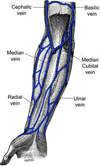
Ulnar artery is NOT an end-artery in case with occluded radial artery. In all cases with occluded radial artery there are documented collaterals, predominantly from anterior interosseous artery. Brachial and femoral arteries are always end arteries. What constitutes acceptable risk of major femoral vascular complications (including retroperitoneal bleeding in perfectly done femoral puncture) in 85 year old lady with ACS and occluded radial arteries: 0.25% 0.125%?

Sunil Rao:
This will be a great debate!! Olivier – make popcorn!

Tak Kwan:
So far Sasko has shown 500 cases without complications. Is anyone know a case of complication of occluded radial and occluded ulnar yet?

Kintur Sanghvi:
I hope I am not annoying any of my radialists friends. Keeping this important interesting debate on……to me the UA is an end-artery if the RA is occluded. Because if UA is not an end artery because of the interosseous branches than brachial is also not an “end artery” as seen in this case from today. Angio and PCI of RCA was performed through the radial recurrent branch in to axillary artery while the brachial is occluded for years from the previous brachial cut down procedure.

Olivier Bertrand:
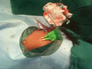
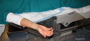
Here is a video and a photo of a patient with occluded right radial and ulnar arteries, we did with Sasko when he taught me transradial carotid stenting. We had to use the left radial after failing to get the radial and we did an injection from the left to prove both occlusions!! (interosseous had grown!) I told Sasko we should publish it as the issue/question would emerge I guess image/video is still worth 1,000 words. Cheers.

James Nolan:
The exact state of the collateral circulation is uncertain in the circumstances of an occlusion of an arm artery. If the femoral is available I can not see why you would not use it, if there is no femoral access the patient is in deep trouble and using an arm artery could be justified. Because Sasko can evaluate these patients and do the procedure safely does not mean the average interventionist can. We should be careful here.

Alejandro Goldsmit:
I vote to make The Debate” in AIM, in one corner…expert favor to use radial and in the other expert corner not agree to use it!!!!

Yves Louvard:
Dear friend, Radial versus femoral in a radial meeting? Are you candidate to support femoral? In UK? (I recommend you not against David Hildick Smith) Ulnar or not when radial is occluded is much better for me.

Josef  Ludwig:
Valentine RJ, Modrall JG, Clagett GP. Hand ischemia after radial artery cannulation. J Am Coll Surg. 2005 Jul;201(1):18-22. BACKGROUND: Hand ischemia is a rare but potentially devastating complication of radial artery cannulation for arterial monitoring. The causes and ultimate outcomes of hand ischemia after radial artery cannulation are unclear. STUDY DESIGN: My colleagues and I reviewed the clinical course of radial cannula-induced arterial thrombosis in eight patients during a recent 5-year period. RESULTS: Mean (+/- SD) duration of radial artery ischemia was 3 +/- 2 days. Injuries were associated with advanced (grade IIb) ischemia that affected the entire hand in four patients and first three digits in the other four patients. Radial artery thrombosis was documented using noninvasive tests or arteriography in all patients. Five injuries were initially treated with thrombectomy and patch angioplasty (n = 4) or vein graft interposition (n = 1); two others were treated nonoperatively with vasodilators, and one was observed without treatment. Three of the four patch angioplasty repairs occluded within 24 hours. Regardless of patency, all patients who survived arterial repairs had continuing ischemia that resulted in digital gangrene or amputation. In contrast, gangrene developed in only one patient treated nonoperatively. CONCLUSIONS: These data show that hand ischemia after radial artery cannulation is associated with high risk of tissue loss or amputation. Operative repair offered no advantage over nonoperative therapy in prevention of digital gangrene in this series. We hypothesize that digital gangrene results from distal embolization from the site of the initial arterial thrombosis, producing ischemia that is not remediated by radial artery revascularization. Nonoperative therapy with vasodilators can be equally effective in treating cannula-induced radial artery injuries in some patients.
And let us be fair. Radial is not a religion; Transcath aortic valve replacement t5he pts are > 85yrs. Last week 105. They all have fem. Access and sheath size far above PCI.

Yves Louvard:
Dear all, I am very sorry not to be at AIM radial this year! (Olivier dont forget me next year) Even if I dont need frequently crossover from radial to another approach, I like the Sasko attitude, I am following it since a long time. Of course radial is not a religion but this is the best way to reduce vascular complications, and the best way to make it true for patients is to adapt the technique to perform 100% radial (+ ulnar ?) I will fail, only 97% today. The method is to collect the datas from the cases without any other solutions, analyse them, present or publish, have a controversy in AIM, find the scientific reasons why it is working or not, select, do more. Sasko, I heard many comments from the beginning of the radial ! I remember radial is an elegant approach for treatment of restenosis (1997) probably around 2000 I lost a radial  vs femoral controversy in Last Frontier in Interventional Cardiology in Texas 90%/10% recommandations are for people who cannot do this (equipment, volume). We have to go further. It was demonstated for many other topics like LM, multivessel, CTO, bifurcations this is one of the responsibility of our community and AIM Radial ! If we follow recommendations (specially recommendations before official ones), we will not move at all ! Fighting with difficulties make us better even with femoral approach (look also at the BCIS database). Skillness and experience are very important as shown in RIVAL SYNTAX so even if I dont perform Ulnar approach Sasko and others are pioneering, let us look at the datas, let us learn with Sasko dont say this is unethical (remember Josef the 2001 paper refused in JACC without review about 1000 transradial primary PCI). I hope to see the 2 presentations and rebuttal. Please Olivier keep them for me!

Josef Ludwig:
Dear all, I disagree with Yves so far the he and me have learned femoral first. Young operators today start with radial. If radial is 100% how to do interventions requiring femoral. Last week we treated a 105 old pt by TAVI. Not good if they start learnin femoral in this severe ill pts.

Yves Louvard:
But Josef, fellows in our group are begining structural after 2 years! of course not from nothing as we did for radial, with a proctor. And as experienced radialists they will become quickly better than pure femoralists even in femoral approach. The futur interventional cardiologists will learn femoral for TAVI like transseptal for atrial appendage closure (are you still doing transseptal Josef ?)! Nice to discuss again Josef.

Josef Ludwig:
No, because of my neck I am more writing than doing cath work. Dear friends, I remember that I was reading some yrs. ago that anatomic variation of the radial are unilateral in the majority if pts. However, I cannot find the Ref. Can anyone help?

James Nolan:
Josef, I dont think it is well documented, but some variation, such as subclavian tortuosity, is definitely more common on the right than the left and the data for this is in the Italian studies of left v right radial I have also seen something suggesting loops etc. are often unilateral but I dont have the reference to hand

Yves Louvard:
Dear Josef, You heard that in Massy radial meeting at the end of the 90s Congratulations Presentations were done by Dr Rodriguez Niedenfuhr (anatomist). Here are my 2 (one two) reference papers by him. You will find that there are no rule for bilaterality of vascular anatomical variations in arm/forearm (thousands of autopsy) Best regards.

Ian Gilchrist:
Here are 3 (onetwo three) anatomic articles that discuss asymmetric arterial patterns.

James Nolan:
Heavy going those articles. So, it looks like about 50% are bilateral, 50% unilateral?

Josef Ludwig:
1 study 38%, <50%. Asians may differ.

Tejas & Sanjay:
One should avoid to puncture ulnar.

Editorial Board Comments:

Tejan Patel:
1) Blisters caused by following.

  • To much pressure
  • Reaction between antiseptic and radial pressure device applied.

2) I use NTG 200-500mcg and Nicardipine 200 mcg.
3) Blisters at Radial artery site is never from Heparin.

I would suggest use minimal pressure at Radial artery site to prevent bleeding and wipe of all the antiseptic before applying radial band this will almost eliminate blister problem. Also try note to use plastic adhesives as a dressing, just simple bandage will work.

Samir Pancholy:
Agree blisters around puncture site are from local reaction to drug or pressure. Heparin is not given locally at puncture site so not likely the cause. Blisters on proximal forearm are different and caused by vasodilation induced mechanism seen especially with Diltiazem.  We use intra-arterial nitrates 200 mug with verapamil 2.5 mg in one lab and 200 mg nitroglycerin with 5 mg of Diltiazem in other lab that I work in.

Mauricio Cohen:

  1. I agree. The blisters are probably related to the high pressure in the compression device.
  2. To reduce radial spasm what radial spasmolytics you are using commonly? I use 3 mg of Verapamil or 300 mcg of Nicardipine. Sometimes I do not use anything.
  3. Is blisters are heparin induced? Heparin is given IV in my lab. I have never seen blisters actually.
Ian Gilchrish:
The only time I saw skin blisters was with the old Hemobands when they were put on too tight.

Ronald Caputo:
I have seen it resulting from swelling and edema related to a hematoma – as above.
I do not think that is a hypersensitivity reaction to the band but if adhesive (tape or an onsite) is used to fix the sheath in place that would be another possibility.

Tejas & Sanjay:
Blisters are due to pressure necrosis by TR band not due to heparin. Patent hemostasis method should minimize the blisters. Early Removal of TR band helps decreasing the incidence of blisters. Inj. NTG 200 microgram / Verapamin 2.5mg / Diltiazem 5 mg are effective to reduce radial spasm

Josef Ludwig:
A blister can develop if the skin is rubbed for a long period or if there is intense rubbing over shorter periods. Friction blisters often occur on the feet and hands, which can rub against shoes and handheld equipment, such as tools or sports equipment. Blisters also form more easily on moist skin and are more likely to occur in warm conditions.

Editorial Board Comments:

Sunil Rao:
No idea. There may be some literature in anatomy journals. I do not think I have ever seen a low origin radial

Ian Gilchrist:
Depends on height which is reflected in arm length. Low bifurcation is not likely due to embryology of radial artery.

Josef Ludwig:
No idea

Tejas & Sanjay:
Length of the normal RA from brachial bifurcation to the head of radius bone varies from patient to patient & roughly it is nearly same as forearm length. We have never  seen low origin of radial artery.

Ian Gilchrist:
Due to the development process thought to be involved in the formation of the radial artery, a low take-off is unlikely. The origin of the radial initially is much higher than in the adult form and thru maturation appears to migrate down to antecubital fossa. Residual forms of this process are represented by the high take-off radial (one that never migrated or partially migrated down) to the remanent accessory radials that represent residual artery structures in the path of the former radial.

Editorial Board Comments:

Sunil Rao:
Chances are that this was a high radial attachment or there was a loop and the catheter was in a recurrent radial artery. I would have switched to the left radial.

Samir Pancholy:
Agree. There was either an issue with anatomy or spasm. The next step would have been to perform an angiogram to delineate the anatomy. Escalating with equipment without anatomy delineation could lead to major complications. Terumo Heartrail catheters have braiding that is different from the rest and so even though they are not hydrophilically coated, they have less friction on the exterior surface. That is why it succeeded but if anatomy was real adverse and spasm was intense you could have had entrapment. Radio brachial angiogram is a must when you encounter resistance.

Kintur Sanghvi:
It is very unlikely to have spasm at the innominate artery. If the obstruction point was in proximal innominate artery (proximal to vertebral origin) more than likely it was an obstruction because of the mismatch profile of the ID with the OD of 0.035 wire. The wire straightens the vessel to some extent and caused bias. Diagnostic catheters have smaller ID AND the mismatch is not as bad. When you came with different shape catheter it changed the bias of the wire by moving the wire just slightly away from the lumen wall. This situation can be rectified by using a softer wire (0.35 glide or v0.18 wire)clocking or counter-clocking torque while advancing the catcher or by using ballon assisted technique.

Josef Ludwig:
I might have probably injected 5-10 times NTG at o.2 mg per injection. If this did not help I would have switched to left

Ronald Caputo:
I agree. Always take a picture when there is trouble advancing a catheter and you were probably in an accessory radial artery originating from the axillary. Spasm or a kink where the artery joined the axillary caused resistance.

Mitchell Krucoff:
It may be that your jr4 guide encountered a plaque in the in nominate. It does not have to be a tight stenosis, but may just be a shelf which can catch the front edge of a guide catheter where the more supple tip of the diagnostics passed easily. Frequently this kind of plaque is calcified and may be very rigid, and if the curvature of the vessel and the guide wire bias combine a guide may stop abruptly.

Key tips might include:

  1. Do not push. The solution here is finesse not force.
  2. Most frequently backing the guide up away from the obstruction and rotating the guide as you put some traction on the guide wire is enough to move the front edge of the guide off of the plaque shelf and into free space. This may be what happened when you changed guides–the tip of the Ikari was different enough to find a new path.
  3. Changing wires may help although a glide may not be the best choice as what you really want is to change the wire bias by changing the wire–a wire like the Terumo Advantage glide has a very stiff body and that would be my next option.

A shelf obstruction like this feels very different than spasm. As you back the guide up a little completely free movement will return as the tip comes off the obstruction. With spasm even after you back the guide up you would feel the dragging continue. Hope this is helpful.

Tejas & Sanjay:
Inject and define the anatomy. Most of the times, it is anomalous RA arising from high brachial or axillary artery. In this situation usually the RA calibre is smaller than normal. Balloon-assisted tracking technique leads to success in most situations.

Editorial Board Comments:

Ian C. Gilchrist:
I am sure others have their own versions as there are a variety of ways to reach the goal and depends somewhat on the tools you have at hand.

  1. Use a .035 wire or similar (not PTCA wire),
  2. Take your catheter in the central aorta and use it to direct the wire towards the left subclavian. You can use anyone of a variety of shapes to point your wire. Everything from a pigtail, to Amplatz, to Judkins, etc. that will redirect wire works at times,
  3. Direct wire down the subclavian into distal arm (brachial level or below). This gives you a rail to work on,
  4. Remove catheter used to direct wire and put on mammary catheter (IM or Judkins right, or other possibilities) on wire. Using small French sizes allows better tracking along wire,
  5. Advance catheter to distal subclavian,
  6. If wire tries to prolapse out, can secure in left arm temporarily with blood pressure cuff inflated or forearm bent,
  7. Once well into subclavian remove wire. Important to be well into subclavian as catheter may recoil somewhat,
  8. Rehook up for angiography,
  9. Slowly pull back with slight counter clockwise rotation to angle the tip of catheter towards anterior chest wall and you should find it.

If catheter falls into aorta, repeat steps above. It seems unnatural at first, but then becomes easy after you understand the steps. Good luck.

Samir Pancholy:
True, many ways to get there. In 40 degrees LAO, I use a TIG catheter, with baby J wire and after entering the descending aorta, “everting” the catheter in the arch to face the left subclavian, advance the 1.5-mm J wire to left subclavian once you either engage it or have a straight shot at it. After enough purchase with the 1.5-mm J, advance the Tiger into subclavian. If difficulty in advancing, use jack-hammer movement of the catheter or bend the left elbow in a jack-knife position to trap the wire and then advance the catheter (Patel T, Patel T et al). JL catheter can be used to replace Tiger to get in subclavian and then exchange for LIMA catheter etc. Once in left subclavian, I engage LIMA in RAO cranial position to lay out LIMA ostium. After parking Tiger in left subclavian distal to LIMA gently counterclock and it will “crawl” back. If you pull it will frequently end up in the aorta. Once again, many ways to get it done.

Olivier F. Bertrand:
Here is our technique as published a while ago OB. (click here)

Ronald Caputo:
I use the exact same technique as described by Olivier except I use an AL-2 to engage the left subclavian. I prolapse it in the descending aorta and pull it back. A non-selective shot is always done prior to exchange for an IMA.

Yves Louvard:
Dear all, I appreciate the paper of Olivier as I reported the first time in PCR course a close technique probably in 99. Some subtle differences:

  1. At the end of coronary angio I take a mammary catheter 5F (preferred Cordis),
  2. I go to LAO 30 degree,
  3. Turn clockwise the catheter to enter horizontal aorta and go to descending 4.  Remove the wire and, pull the catheter pointing up, it will never enter the left subclavian. Injection to locate the subclavian, if not seen a small counterclock,
  4. When seen I use a Terumo “shapeable” with a J and a slightly longer shape proximally. The “J” only frequently go to vertebral or other branches and push back the catheter in ascending aorta,
  5. Very important as said by Olivier to push deeply the wire, in arm or even forearm (with drilling…),
  6. When the wire is down, push the catheter better with a clockwise rotation distal to mammary,
  7. AP projection, pull the catheter to the begining of post vertebral subclavian,
  8. Remove the wire, it is stable!
  9. Then, counterclock (do not pull) to move back the tip (in the mammary), if you are proximal turn clockwise, do not push, to move forward.

Additional tips:

  • To bock the wire, close the elbow or inflate a cuff (no nurse under X-Ray),
  • In case of failure a Simoons 2 can be used to enter the subclavian, but maneuver is a bit more complex, and it is necessary to exchange on a deep waire for a mammary catheter in a high percentage of cases,
  • I tested two Yumiko (first name of a female Japanese colleague I encounter in Saito meeting: “Crazy of the Radial” in Kamakura in  2000!) coming from Japan, probably the best option, but impossible to get in France,
  • I agree for 5 french only PCI for deep intubation and support (on a very distal 0,014 wire of course).

Most recent success rate in 2012-13: 93.3% selective / subselective with good diagnostic quality: mammary graft and distal branches. Try first in younger patients (less calcified), no long lasting HTN (less sinuous) … It works!

David Hildick-Smith:
Dear Dr. Abdul, At the risk of offending my colleagues on TransradialWORLD I would suggest that the correct way to approach the LIMA is usually from the left radial approach, or, in cases of bilateral IMA, from the FEMORAL artery (shock, horror). It is challenging, and fun, to catheterise the LIMA from the RRA, and we all have examples and descriptions, but that does not necessarily make it the safest or most sensible approach. Radialism is not a religion, its just the best way to the heart in most cases. There are exceptions though, and bilateral IMA is one of them!

Tejan Patel:
With Terumo Baby J wire (1.5mm) now LIMA engagement have become much easier and with less radiation also. You can use any catheter that points upwards (Tiger, Jacky, AL1, 2, 3 or even LIMA) and just advance Baby J wire and invariably it will go to Lt. Subclavian and advance until wire is in Left radial/Ulnar artery and any 4 or 5 F catheter will track over it. All the suggestions on this site will work and you will just have to figure out yourself what works best for you. I use either Tiger or Jacky and 4F Cordis LIMA and it works 90% of the time with selective engagement of all the grafts including LIMA/RIMA. With Baby J Wire our routine Fluoro time is around 3-4 minutes for last 2 years. If needs PCI best approach after you have master this technique use LIMA guide and most of the time you can finish the case with some patience. More advance technics will be published in near future with the advanced Tips and Tricks atlas by Tejas and colleagues. You can also refer to various tricks published by Dr. Sasko Kedev. How to cannulate cranial vessel from ipsilateral and contralateral approach. Best of luck.

Olivier F. Bertrand:
VADE RETRO SATANAS!!! 🙂

Sasko Kedav:
We routinely cannulate left subclavian with Simmons 2 catheter after reshaping it with wire in the descendant aorta. Usually it provides good imaging of LIMA. If not, we wire the subclavian and exchange with IM catheter in a manner already described. For LIMA PCI we use the left radial access. For cannulation of left common carotid with Sim 2 already in subclavian, just push, conterclock rotate and pulled back in left common carotid.

Mauricio G. Cohen:
I agree with Dave. We have all engaged a LIMA from right radial approach and a RIMA from left radial approach. However, I do not think that this practice is safe because of the degree of manipulation and the additional fluoroscopy time needed. The best way to approach an internal mammary artery is through ipsilateral radial approach. Because of the origin of the RIMA from the vertical part of the right subclavian, using a regular IM catheter may be challenging. We have successfully used the VB1 catheter in those instances. In cases of bilateral IMA grafts, bilateral radial access is probably safer than transfemoral. See attached paper that has a section on LIMA / RIMA access. I hope this helps. Best regards.

Mitchell W. Krucoff:
Agree with many that left radial is ideal for LIMA, if the surgeons do not take it out as a graft conduit! With practice, right radial is very useful, and RIMA and LIMA both accessible. I use an IMA diagnostic to cannulate the LIMA, then pull back through the innominate to the right subclavian and shoot the RIMA. I actually like the Terumo Advantage wire best for crossover from right radial to left subclavian. Ian gilchrist introduced me to this wonderful wire several years ago. Manufactured originally for peripheral cases, it has a steerable Glide tip, very robust wire body, so no prolapsing into aorta when exchanging to IMA catheter. Even an IMA guide catheter goes easily over this wire. When using the Advantage, as mentioned by others any up-pointing catheter works TIGR, Left Judkins, and Amplatz.

Tejas & Sanjay:
We developed a technique (click here) for cross-over from right subclavian to left subclavian artery during RRA, which was published in CCI a few years ago. It not only helps cannulation of LIMA but also helps doing left subclavian, left vertebral & left ICA intervention. It is attached herewith. Best regards.

Editorial Board Comments:

Ian Gilchrist:
Never seen that in 20 years. It has been a long winter in the Rochester area of the east coast (cabin fever?)

Rajiv Gulati:
Did it happen on April 1st Tejan?

Samir Pancholy:
Agree, never heard of it. Likely unrelated, supratentorial.

Tejan Patel:
It sounds weird but it happened. We tried multiple time and it will happen every time we put the band.

Ronald Caputo:
Did you plug that band into an outlet? Just kidding. Sounds like either a subjective response or myoclonic reflex.

Tejas & Sanjay:
We do not have VascBand available in India.

Editorial Board Comments:

Sunil Rao:
Thank you for your question. I am not sure that I understand it completely, but the vast majority of our transradial interventions are 6-French. We have no problem addressing bifurcations, including unprotected left main bifurcations, through 6-French guides. Almost all manufacturers make non-compliant balloons that allow for kissing balloon inflations through a 6-French (internal luminal diameter 0.070) guide. As long as one is meticulous about not wrapping guide wires, there is not a problem.

Mitchell Krucoff:
Use of multiple guide wires is critical whether you are using 5, 6 or 7 french guide catheters. If you allow the wires to wrap around one another they can bind balloon and stents and make them impossible to deliver. If you get mixed up on which wire is which you can pull one out or advance by mistake and lose access to the position, perforate the vessel, or make other bad things happen. So two things to keep track of: (1) Use of torque in positioning the second wire: After placing the first guide wire, be very aware with the second wire to torque back and forth (clockwise and counterclockwise) do not just go around and around in the same direction. This will help avoid wrapping the second wire around the first one, and avoid trapping the path for balloon or stent catheter. (2) Marking which wire is which: Say you have wires in the LAD and LCX for a left main bifurcation lesion. Several tricks may help you keep track of which is which: (a). Use a short wire for one and a long wire for the other. (b). Use two short wires and pierce a dry 4X4 gauze with the back end of the wire you are not interested in as you go step by step to keep track of which wire is which. (c). Use a folded sterile towel between the two wires on the table to separate the two wires, so that when you fold it up or down there is only one wire exposed for you to work with again, keeping track of double or triple wire settings and avoiding wrapping one around the other are important techniques regardless of guide size or access site. I hope this addresses at least part of your question.

Ian C. Gilchrist:
I would agree with Sunil. The important issue is that not all 6F guides have the same internal diameter and different balloons/stents/wires have different sizes depending on manufacturer and coatings. Depending on your available devices, there may be a combination that will work better than others within the restraints of a 6F guide.

Samir Pancholy:
Agree, except for simultaneous kissing stents everything related to bifurcation PCI can be achieved using 0.071″ I.D 6 French guide catheter. If need be, a 7 French system could be used especially if ultrasound of radial shows a big enough artery. Separating multiple guide wires is a matter of procedural technique and discipline. Yves Louvard demonstrates it the best in live cases, with opposing torque, shaft separation etc. If still have trouble, vascular solution makes a plastic “caddy” to keep the wires organized.

Alejandro Goldsmit:
Thanks for your question, but some PCI would be make by 5 or 6 Fr, 7 Fr are not very common. 7 Fr, could be use by TRI without any problema in CTO, Kissing, etc. It is very important before to start to know the REAL internal lumen of your guied.

Josef Ludwig:
You can do all you need with 6 French even small size Rotablator.

Tejas & Sanjay:
In the situation when you have to use multiple guide-wires in coronary system, particularly in bifurcation stenting, one should first wire the artery which has more complex anatomy as multiple tourques and rotations of the wire should not make any difference. Once it is done, second wire can be deployed in the artery with simple anatomy without many rotations. This can avoid looping of the wires.

Editorial Board Comments:

Tejan Patel:
Best way to deal with Radial artery hemostasis is to avoid any hematoma and still keep ante grade flow all the time! Remove pressure dressing earliest possible. If one has manpower manual pressure will be best making sure you have pulse proximal and distal to insertion site.

John Coppola:
The idea of patent hemostasis is to be able to compress the radial artery enough to stop bleeding but allow flow. This is done by placing a compression device on the puncture site inflating or tightening the device and remove the introducer. The ulnar artery is compressed while looking at a pulse ox tracing. The device is slowly deflated until a tracing appears or bleeding occurs. If bleeding occurs 1 cc of air is placed back in the device. About 3/4 of patients can have hemostasis and signs of perfusion. The minimal pressure method does away with checking for distal flow. The device is deflated until bleeding is seen at which point 1 cc is re inserted. If distal perfusion has occurred this is patent hemostasis if no radial flow this is the best you can do. The idea is to return to the patient and attempt to reduce pressure after 15 minutes, and to continue to re evaluate every 15 minutes with the goal of removal of the compression device as soon as possible.

Samir Pancholy:
I am unclear as to what the exact technique of “minimal pressure hemostasis” is, and how is it different from patent hemostasis. In general, while using patent hemostasis, we use the least necessary pressure to maintain hemostasis, and monitor the adequacy of hemostasis every 15 minutes and readjust compression (if radial not patent) to minimum necessary pressure required to maintain hemostasis.

Josef Ludwig:
How to look after every 15 min in a high volume center?

Olivier Bertrand:
That is a very valid observation. In fact, many centres, even in US, claim that they do patent-hemostasis while in fact they simply use 1- TR-band, 2- with minimal pressure and they do not check the oxymetry while compressing the ulnar When challenged about this modification to original technique, they usually claim that with their volume they cannot check every 15 min while compressing the ulnar. By the way, I believe efforts for standardization remain to be done among us. We do not agree on common defintion of RAO, nor on technique (oxymetry or duplex US) nor timing to assess RAO. In fact, almost all US centers I have discussed with, claim < 1% RAO and OUS, it is 5-15% at hospital discharge. The issue is that most US data come from VA experience which by definition include only males ! just my 2 cents (to quote Mitch)

Samir Pancholy:
I believe checking at a given frequency is a matter of setting nursing priorities. It takes < 7 min of low intensity work for nurses (including charting) to accomplish this (we measured this when our staff rebelled). Just like we have decided to check femoral access site every so often to make sure hypotension is not the first manifestation of the problem, this should be a part of their “jobs” for the patients sake. High Volume or not. I agree with Olivier that RAO needs to be defined (including setting a time domain). Keeping in mind the point of care equipment availability.

Tejas & Sanjay:
The steps of Patent hemostasis technique have already been described in detailed in the literature. Sam Pancholy is the pioneer of this concept. We religiously follow this technique.

Editorial Board Comments:

Yves Louvard:
Empirically what we do in Massy is inflation with 15 cc, followed by 2 cc deflation every 15 minutes after angio (5000UI heparin) or every 20 minutes after PCI (heparin enough for > 300 sec ACT). Normally after the first (or second) deflation we have an antegrade flow.

Mitchell Krucoff:
We take advantage of the visibility thru the TR band and do not check ACTs at all. At any stage if there is bleeding the nursing orders are to put air back into the TR band to stop the bleeding and then re-start the clock. For a diagnostic procedure (we give around 5000u heparin in full size patients, about 70u/kg): we wait 30 minutes after sheath is pulled, then remove 3 cc of air every 15 minutes. For a PCI (we often use bivalirudin) we wait 90 minutes after sheath is pulled and then begin the same regimen. If a patient is on coumadin we do this the same way regardless of INR. If bleeding occurs, the TR is reinstalled until it stops and the clock starts over. ONE NOTE: It should be appreciated that while we use standard orders, a 3 cc of air is not the same for every case. In achieving patent hemostasis we try to leave at least 10 cc of air in the TR band, but the pressure/volume loop of the TR band is also strongly driven by how tightly you fasten the Velcro band itself. If you put it on too tightly, you may only leave 5-6 cc of air to have patent hemostasis, and then nurses taking out 3cc at a time essentially are just taking the device off. My 2 cents (or 3cc, whatever) 🙂

Tejas & Sanjay:
Our Strategy is to reduce 1 cc air every half hourly. We remove TR band, 4 hours after the procedure.

Editorial Board Comments:

Mauricio Cohen:
Hi Tejan, I really like the small radius wire or so-called “baby-J”. My impression is that it does not go into side-branches as the angled-tip glidewire and it does not cause much spasm as the regular J wire. In addition the regular J sometimes gets stuck in the vasculature because the J is too large for a 2-3 mm vessel. I use the baby-J wire routinely and feel reassured that I wont cause harm. I usually advance without fluoroscopy until I feel the slightest resistance, by then the wire is already in the aortic arch. I think that this strategy saves fluoro time. Merit Medical also makes a non-hydrophilic coated baby-J wire. Just my humble comments.

Samir Pancholy:
Agree with Mauricio. Good wire for forearm navigation without fluoroscopy through difficult terrain. Stays out of accessory radials with good tactile feel. The only negatives are for some reason it has a tendency to enter carotid with higher probability compared to the 3 mm J. Also the tip straightening tool is cumbersome and only could be back loaded.

Kintur Sanghvi:
Agree with both the comments. Only reason did not adopt it as a preferred wire because straightening the wire is not possible and I use Judkins curves.

Tejas & Sanjay:
We find it very useful, in certain subsets like Radial/ Brachial tortuosity, Small caliber Radial, Radial/ Brachial loop. PTCA wire requirement in above subsets is minimized.

Editorial Board Comments:

Ian C. Gilchrist:
Short answer is no contraindication. But some of these patients have received extensive instructions not to have anything in ipsolateral arm that emotionally they will request the procedure in the other arm. Not really worth an argument on the table.

Yves Louvard:
Dear friends, I did two radial approaches on the side of a mastectomy this week. There is no contraindication. But it is sometimes a sensible problem with the patient … Explain that you are using the artery, not the vein, if OK do it, nothing will occur … If discussed use the other side.

Tejan Patel:
Make sure no hematoma or perforation in the arm.

Samir Pancholy:
No major issue with radial artery access. Venous access logically not as safe. I would avoid patients treated for lymphedema in the past. We had one case where lymphedema that had resolved a few years ago, recurred after radial “arterial” Cath. Although one should not practice based on anecdotes, it ruined my “clean” record. I have performed about 70+ other cases before that case with no complications. I know Ian has put together a larger series with no major complications. We now calculate the patients bleeding risk using one of the published “score”. If patient has high bleeding risk, radial artery puncture is justified. If low bleeding risk, may be it is better to avoid it. Of course, some patients emotionally will not agree, in which case femoral is the next best option. Overall it is safer probably compared to femoral access in a patient at risk for access site bleeding.

Kintur Sanghvi:
I agree with all the previous comments. Having said that lymph edema is a dreadful complication. The suffering is so much on patient is part that anecdote are enough to drive my decision. I avoid using effected arm radial except in compelling circumstances. Operator in us should be even more careful. It would be very easy to convince a jury that the doctor was negligent….despite expert comments by Ian in the court.

John Coppola:
Having a wife who is a breast cancer survior I agree with Ian that at times there is a large emotional component and despite the fact that it is safe to use the artery it is often better to use the other wrist.

Ronald Caputo:
Arterial cath is safe. Would not do a right heart from that arm however.  The difference between venous and arterial access must be explained to the patient. I agree with John that if there is significant anxiety another arterial approach is an option.

Ian C. Gilchrist:
The flip side (since I was mentioned as an expert witness) is that if a patient s/p breast cancer undergoes a femoral procedure and has a major complication without being offered a radial procedure, it would be hard to defend without evidence that there is a true risk to outway risk of femoral puncture.

Josef Ludwig:
Completely agree with Ian.

Mitchell Krucoff:
Agree with the others, this is a personal/emotional issue, not an anatomic one.

Tejas & Sanjay:
Radial artery access can be used without any problem.

Josef Ludwig:
Radial artery access can be used without any problem???? Look @ picture I sent you all (click).

David Hildick-Smith:
What is the mechanism Josef ? PS Bet it looked like that before the procedure too!!

Editorial Board Comments:

Tejan Patel:
Reassurance and overtime will get better.

Samir Pancholy:
Exercise with extension and flexion of the thumb liberally. Reassurance.

Ian C. Gilchrist:
Time will heal it. Exercise. It will resolve.

Sunil Rao:
It sounds more like some kind of compression nerve trauma rather than vascular occlusion. Should get better with time.

Ronald Caputo:
Bumped the thenar cutaneous nerve. It well get better but I think this points out that radial access is being obtained too distal in the wrist. Prep and stick 2-3 cm higher.

Mitchell Krucoff:
I agree with all that conservative approach and supportive rehab will cure.

Jennifer A. Tremmel:
Nerves can take awhile to recover. Hopefully it will be quicker, but I would prepare your patient for weeks to months, not days, or they will keep bugging you if its not better right away.

Rajiv Gulati:
Yes, I recollect a presented (not sure if published) series of 10 or so pts with superficial radial nerve sensory neuropathy, some of whom had abnormal nerve conduction studies. All pts had resolution of symptoms by 3 mths. Agree with reassurance. Exercise also make sense. Found link to the poster (click here)

Tejan Patel:
Any role of lyrica or Gabapentine?

Olivier F. Bertrand:
Great thx for sharing. I met a patient recently who has had more than 12 repeat right radial….Last time prior his cath, he mentioned to me that during his previous attempt, the resident had tried very distal and he could feel the unsuccesfull attempts… At that time he showed severe thenar atrophy and had become unable to touch the little finger with his thumb….I took pictures and intend to report it.

Josef Ludwig:
I really very surprised by the expert comments around the globe. We in Erlangen – and I looked up yesterday for preparing a lecture – we do 1.500 rad PCI and about 2-3.000 angio per year. In 10 yrs this amounts to 30-40.000 procedure. Noone  here in little Erlangen ever observed this problem. Thus, it MUST be a very rare complication. Wherefrom is thus coming all the “evidence-based” expert comments??? I wonder and wish you all wonderful day.

David Hildick-Smith:
I have not come across this problem and I would agree that it seems likely that the approach is too distal. Distal approaches to the radial artery have, of course, the additional disadvantage that vasovagal reactions are commoner. Go a few cms above the styloid process and this should not occur.

Tejas & Sanjay:
Reassurance. It will disappear with time.

Editorial Board Comments:

Josef Ludwig:
I have heard about this before. Fortunately, never have been forced to this problem, but what I was told is to wait and not go to surgery. And, this is acceptable because what surgery want to do with an occlusion on radial. What I was informed on this very rare Situation is to ask a neurologist or pain speziell. Cool and anti-immflammatory med such as Arcoxxia.

Ian C. Gilchrist:
Unless there is overt arm ischemia, I would not give a surgeon a chance. Without overt ischemia, it is neuropathic or pain from arteritis that will resolve with time. Surgical manipulation (except for true compartment syndrome) will only increase long term problems. Attempts at re-establishing flow with surgery is not helpful and should not be done in most circumstances. The surgical outcome in the literature to revasc a radial artery almost always ends with a chronic occlusion. Warm compresses, antiplatelet therapy and hand exercise to encourage blood flow along with non-steroidal anti-inflammatory agents almost always work. Good luck.

Sunil Rao:
Agree. Conservative management is indicated.

Mauricio G. Cohen:
We have all seen this picture before as it occurs in a very small proportion of patients after TRA. Conservative management is indicated with reassurance to the patient. A surgeon may indicate surgery (is not that obvious) or prescribe oral anticoagulants. I agree with Ian and Sunil. Sometimes a short course of steroids may help. Good luck with your patient.

David Kandzari:
Agree with others. I have had success with short course of tapering steroids.

Tejan Patel:
In my experience time is the best healer. Any surgical Intervention will be of no benefit. For pain control use pain meds, NSAID. Short course of Steroid/Medrol dose pack might help shorten days of painful hand. As I have mentioned before on this site “Ischemic Preconditioning” will help. (Use manual BP cuff and inflate it to occlude blood flow in the affected hand for 2-3 minute and release for 30 seconds, repeat it few more times) in my limited experience I have seen dramatic improvement. I wonder anybody have tried Highdose Viagra?

Sunil Rao:
I think Kandzari and Caputo have tried high dose Viagra, but for a different indication!!

Samir Pancholy:
Agree with it likely being most likely arteritis and treating it with mentioned approaches. In the absence of forearm hematoma, surgical involvement will be redundant. Because it seems the last catheter withdrawal was painful, I am assuming integrity of brachial ulnar vascular status was confirmed.

David Kandzari:
Ha! I was thinking the same thing! Maybe we just did not have the right dose!

John Coppola:
Agree with conservative approach. I have use tapering dose prednisone and small dose 300 of gabapentin at bedtime for only patient generally warm soaks and nsai work.

Ian C. Gilchrist:
Clarification. Tejan, in your experience is the Viagra to pass the time or does it relieve spasm in the radial artery?

Tejan Patel:
Ian, for both, I think increased Nitrous Oxide might be of help to relieve neuropathy and pain.

Alejandro Goldsmit:
I am agree with conservative approach, but anticoagulation, will be very helpfull Viagra, is a good option, but only in diabetic patient. Handgrip manuver and warm pads over the arm are a good tools to help, plus any pain killer pils and the most important is not waist time in expensive and unusual test. In 20 to 30 days, is very common to disapper all the simptoms.

Mitchell W. Krucoff:
I agree with the previous comments, but would it seems worth clarifying that there may be two processes going on:  occlusion of the radial artery, and arteritis around the level of the elbow. Presuming the cath was done with 5 Fr or even 6 Fr diagnostic catheters, feeling them on removal suggests spasm during the procedure, which could contribute to both. The arteritis is absolutely not a surgical issue, and will resolve with time and possibly non-steroidal anti-inflammatory drugs. The radial occlusion in itself sounds asymptomatic. As comments have already mentioned, surgery is more likely to add troubles not solutions. If conservative management does not recanalize, Sam Pancholy and others have reported percutaneous recanalization if the benefit/risk warrants, that would be something to consider.

Ronald  Caputo:
Completely agree with all comments.

Tejas & Sanjay:
Radial artery occlusion is not responsible for pain in elbow crease. It seems like neuropathic pain. Reassurance and analgesic medicines should help.

Josef Ludwig:
I completly agree, but, none the less, of course caused by radial cannullation.

Editorial Board Comments:

Mitchell Krucoff:
We generally do not interrupt coumadin. If the INR is therapeutic, we do tell the patient that if we cannot complete the procedure from right or left wrist that we will not be going to the leg, and patients accept this very well. Super-therapeutic INR I usually weigh against the acuity of the cardiac presentation. For ACS I would just go ahead based on risk/benefit. For chronic presentation we might look into why the INR is too high and re-visit timing of cath, again based on risk/benefit of adding heparin plus more intensive anti-thrombotics on top of coumadin if PCI is performed.

Samir Pancholy:
We do not stop warfarin. We do the procedure if INR < 4. Give 50 U/Kg heparin for diagnostic catheterization like we always do.

Ian C. Gilchrist:
I do not stop coumadin or any of the new oral agents. There is a growing body of literature showing both thrombotic risk to stopping oral agents and likewise a bleeding risk to most of the various bridging approaches. For elective cases as long as they are therapeutic I continue with procedure and add heparin on top. In emergent cases, I have no fixed INR limit but do decrease heparin dose if INR is high. Bigger question is what to do if you do an intervention and which drugs to use (triple therapy?) and which stents. The actual procedure with regard to coumadin is the easy part. Also in the end, you do not need to restart the coumadin if you never stopped it. Makes life easier.

Josef Ludwig:
Ian, do we need triple therapy? If we need coumadin just implant bare metal and coumadin and clopidogrel will do instent-restenosis is like surgeon causing celloid, but saved life! Long-term restonis is another animal. With good weapons to fight.

Kimberly Skelding:
Never stop coumadin unless I am planning on doing a CTO and of course then the anatomy is known. I also give heparin as well. I did not do that in the past and had a thrombotic event in the arm. I have never had one since I continued to utilize heparin.

Kintur Sanghvi:
I understand that we are not concern of the INR with radial access. And the notion that I only avoid CTOs with high INR may give a wrong message. We lost a patient in 2005 during my cardiology fellowship from wire perforation while the INR was 2.8 or so. We were the new Radial center and became careless about the INR. From that memory I do not perform planned intervention with INR higher than 1.6 unless for STEMI or refractory unstable angina. I do not stop anticoagulation for diagnostic procedure but do not perform an add-hoc intervention with high INR. Two weeks ago I had a big mushroom perforation with a NC balloon rupture in a calcified mid RCA. That patient I had stopped one of the newer anticoagulant two days before. It could have been a different nightmare, if I did not. Patient did fine and required a graft master. I remember Johns (Coppola) saying, “If you have not seen it yet, you have not done enough”. Radial access is for patient safety and I do not see a reason to put patient at unnecessary risk for elective interventions.

Tejas & Sanjay:
We do not stop warfarin for diagnostic or interventional procedures, unless PT-INR is > 4.0

Editorial Board Comments:

Ian C. Gilchrist:
“Absolute contraindication is a bit harsh, perhaps a conditional” contraindication. I think that the condition needs to be respected, but can be managed successfully if treated expectedly. Use small catheters, sedation, reasonable anti-spasm regimen and do not let the hand get cold. It is better than a retroperitoneal bleed. On the other hand, the patient also needs to be aware of potential issues and be part of the discussion. So far, the literature has not suggested a major risk although there are clearly some antidotal experiences around. Just my personal thoughts.

Jennifer A. Tremmel:
I agree with Ian. There is a spectrum of Raynaud, from mild (isolated, occurs with extreme cold) to severe (associated with a rheum disorder and tips of digits can be lost). The more mild forms can be done with proper technique as mentioned by Ian. The more extreme ones I might avoid, but fortunately they are rare.

Yves Louvard:
I have performed several times (5?)  transradial approach in Reynauds patients. I recently … No problem but small population, no systematic attempt, no specific follow up … Prospective registry?

Ronald Caputo:
Absolute is often in the eye of the beholder. There are varying degrees of Raynauds. There are cases where TF is more dangerous or not possible. The severity of the Raynauds as well as the need for TR approach must be balanced. Generally I take no chances when the well being of a hand (especially dominant) is in question. Why ask for trouble?

Mauricio G. Cohen:
In general I agree with the posted comments. Radial is not a religion. I recommend following the general rules that we apply in all cases:

  • Assess radial artery diameter to anticipate spasm/ risk of occlusion,
  • Use the smallest sheath/catheter diameter possible, &
  • Make every effort to prevent radial artery occlusion.

Kiemeneij already said that Radial is not a religion. Judgment is important. I hope this help.

Yves Louvard:
But he remains the pope! More seriously. The way the radial approched progressed these last 20 years was related to limits. Each we faced a problem each time we tried to find a solution, sometime we succeeded, sometimes not:  in my center renal failure is a contraindication to femoral, I control left mammary artery from right radial. So, radial is not a religion, but a solution with some remaining challenges. We have now a lot of enthousiastic young operators to face these challenges.

Samir Pancholy:
I personally avoid patients with systemic sclerosis (CREST) with severe Raynauds completely. Three cases that have created a very unpleasant memory with spasm/ ischemia of the hand in all three cases to a variable degree, taking hours to resolve with IA meds initially and then IV meds. Milder cases of temperature related digital vasoactive complaints in patients with no aggressive systemic disorder, do well with no consequence in our limited experience. Fortunately not a common problem.

Mitchell W. Krucoff:
I agree with most of these comments: clinical judgement must be applied. If someone has severe Reynauds, may also be associated with migraines and even coronary spasm, If might go femoral. If it is less severe and, for instance, the patient is obese, I would go radial but might give additional local intra-arterial nicardipine with verapamil.

Josef Ludwig:
I really do not know, but, I myself would refrain doing TRI in this disease.

Tejan Patel:
I would avoid TRI in these conditions.

Tejas & Sanjay:
Yes, however, we rarely get such cases.

Editorial Board Comments:

Mitchell W. Krucoff:
In our lab: 1. We use 2.5-3.0 mg verapamil into the artery immediately after access and the again immediately before pulling the sheath. We give it slowly and mixed with blood to avoid stinging, but have not seen heart block or hypotension. 200 micrograms nitro glycerine also works, but is shorter acting and may cause some hypotension. 2. We give heparin via the IV once we have a wire up to the aorta.

Ian C. Gilchrist:
While Dilt and Verapamil do not usually contribute to bradycardia, they potentially can. Nicardipene and some others calcium channel like blockers found outside of US have no effect on conduction and can be used regardless of underlying rhythm. All the calcium channel blockers have relatively similar half-lives (much longer than NTG) and vascular effects, they differ on their effects on the conduction system.

Samir Pancholy:
We use nitro 200 mcg + 5 mg of Diltiazem ia. Verapamil 2.5-5 mg could be used Sinus bradycardia is not an issue although if AV block (>1st degree) will use nicardipine. UFH we give IV.

Tejan Patel :
I still give all the meds IA. Heparin 3000, Nicardipine 200 mcg. And NTG 200-400 mcg. Depending upon BP. Always use 0.9 NS 300-500 cc bolus just before and during the procedure to avoid hypotension. For few years now no issue of spasm or any hemodynamic effects. We try to keep pt. Comfortable without sedation.

Kintur Sanghvi:
Radial perforation is extremely rare. That is not the reason to give Iv heparin. In most cases of radial perforation we like to  give heparin. It is to avoid burning in RA that we give iv heparin.

Alejandro Goldsmit:
Dear Ajay, recently with Dr. Tejas Patel, and other coauthors, published a registry to know real incidence to spasm, and severals strategy to avoid it. Click here

Mauricio G. Cohen:
I give heparin after have I reached the root of the aorta. The reason is not fear of radial perforation, but to make sure that I can complete the procedure from transradial access. When I reach the root of the aorta, I am 95% certain I do not need to crossover. It is not a good idea to have the fully anticoagulated if you need to crossover to femoral access. Even though this is a very rare occurrence (< 5% in the literature), femoral hemostasis is more complicated in the anticoagulated patient. You need to wait 2-4 hs to remove the sheath manually affecting the throughput in the holding area. Preferred cocktail is a calcium channel blocker (Nicardipin 300 mcg or Verapamil 3 mg) through the sidearm of the sheath. I hope this helps,

Tejas & Sanjay:
We use only NTG (200 mcg) as a cocktail. Dilzem & Verapamil should be avoid in presence of bradycardia. Inj. Heparin should be given immediately after sheath insertion. If perforation occurs then, it can be reverted with Inj. Protamine.

Editorial Board Comments:

Ian C. Gilchrist:
We have the nurses give the heparin.

  1. It avoids pain in the artery. Pain = something bad (not sure exactly what), Just as effective IV or IA,
  2. Avoids having the doctor forget to give heparin,
  3. Engages the staff in the case as they know it is their job to give the heparin and remind the doctors if they have not been given the order,
  4. Is a great task to put on a check list for the procedure,

Good leader keeps 10 people working, does not do the work of 10. Get your whole team on board! (my opinion)

Kintur Sanghvi:
Thank you Dr. Ian, For the leadership lesson.

Jennifer Tremmel:
Our nurses put 100 nitro, 2.5 verapamil, and 5000 heparin into a 30cc syringe. Once we place the sheath, we attach the cocktail syringe, fill the remaining cc with blood, and then inject it into the arm. Sometimes it burns a little, but we never have spasm. This is how John Coppola taught me years ago, and I still do it this way. There is no right or wrong way to do it as long as it is done. You just need a consistent system, and I am sure this forum will give you about a hundred to choose from 🙂

Yves Louvard:
For us it is close to the Jennifer Tremmel way: 10 cc syringe is attached to the sheath with nicardipine and 5000 ui of heparin which is slightly burning. No spasm.

Samir Pancholy:
We give unfractionated heparin intravenously as it burns less IV and RAO rates are equal (AJC Oct. 2009). Better for public relations amongst the whiny Pennsylvanians. Although there is no difference in efficacy, the Gilchrist argument of “team” effect is I believe huge. Our nurses love the fact that they “control” heparin end of the cocktail. Makes them feel the spirit of pride and responsibility. We have unpublished data (quantitative radial angiogram) that show that UFH is actually a very mild vasodilator when administered as a bolus. We can talk more about this if anyone interested. There are some basic science reports corroborating local endothelial dependent NO release by giving bolus heparin (which is why it might be causing some of the burning, very much like other vasodilators). So spasm has never been an issue with IA UFH in the past.

John Coppola:
I have  nurses give It is  less painful just as effective dose 50units per kg to max 5000 units. Use I have heparin found it is less painful have nurses give once I am in ascending aorta at dose of 50u/kg.

Mauricio Cohen:
Dear All, I use the same approach as John.

  1. Route: Intravenous
  2. Dose: 50 U/Kg –> Max: 5000 U. Transition +/- to bivalirudin if PCI.

One thing that I do different is that I give heparin IV after I reach the ascending aorta with the catheter and know I will be able to engage the coronaries. It is more complicated to crossover to femoral (after trying the contralateral arm) in a fully anticoagulated patient.

David Hildick-Smith:
Heparin is acidic. It is therefore painful injected in the radial artery. Give it in the aortic root.

Tejan Patel:
To avoid clot- We give Heparin 3000 -5000 always IA (always dilute with blood, always warn pt. it might burn for few seconds). We always use pressure manifold and flush with Heparinized 0.9 Saline and making sure have proper Arterial Wave form with sheath. I believe IA is better than IV, reason mentioned (acidic, negatively charged, avoids first pass effect and probably more local effect compare to IV and also vasodilator) We almost never sedate pt. with this approach most of the pt. does not complain. We always use long Terumo sheath and despite that with above approach in more than 10,000 radial cases had no clot issue. To avoid spasm- We start with bolus of 400-500 cc of IV Saline. Heparin as above, Nicardipine 200 mcg. and NTG 200 mcg all IA. With this cocktail always use counter puncture tech. with angiocath needle with plastic cannula. With this approach we have less than 2% of Radial conversion rate even with CABG pt.

Ronald Caputo:
I give it in the aortic root but it can also be given IV by the nurse.

Alejandro Goldsmit:
Dear team, we shuold use heparin IV, slowly and no spasm, no painful. After start the procedure, 4 ml nalbuphine, to avoid spasm, NEVER use other cocktail.

Mitchell W. Krucoff:
we give peripheral iv for patient comfort

Kimberly Skelding:
I always use IV and generally 5K.

Josef Ludwig:
We administer 5,000 U i.v for diagnosis and 10,000 U for PCI and, if longer procedure, we measure ACT every h.

Tejas & Sanjay:
Our routine practice is to give I.V. Heparin. I.A. Heparin many times produce pain and burning sensation. This may precipitate vasovagal episode. Pharmacologically efficacy of I.V. & I.V. heparin is the same.

Tak Kwan:
I give 3000 IV now because of small size of my patients.

Editorial Board Comments:

Sunil V. Rao:
Our protocol is the following: Place hemostasis device, ensure patent hemostasis – wait 30 min if diagnostic, 90 min if PCI – release 3 cc of air every 15 minutes (NOT over 15 min, but release the 3 cc all at once, then wait 15 min and then release another 3 cc, etc) until empty. It has worked well for us for the past 6 years with very low incidence of re-bleeding. The re-bleeds we see almost always occur in patients with therapeutic INRs.

Mitchell W. Krucoff:
One aspect of this protocol: If there is bleeding the orders instruct to put 3cc back into the band and re-start the clock on the whole process. This protocol also works nicely with patients on coumadin with therapeutic INRs. I do not know of any data comparing any two deflation protocols head to head.

Tejan Patel:
In my practice, Diagnostic cases we start removing 2 cc at 30 min and dressing at 60 min. Interventional cases start removing 2 cc at 60 min and dressing at 120 min. If Pt on Coumadin with INR more than 3 than only I follow Interventional protocol otherwise still follow Diagnostic protocol. Patent hemostasis is best practice. Beyond 60 min. In diagnostic and 120 min in Interventional cases MANUAL pressure with still patent hemostasis will be the best thing. We all need your (RN and recovery unit staff) help to prevent bleeding at site and still keep Radial artery patent to perform another endovascular procedure some other time. There is no better compression device than Proper Manual pressure.

Kintur Sanghvi:
Dr. Pancholy, Awaiting your response. From my understanding the TR Band looses air over time on it is own (I do not know if it was designed with that purpose). Please correct me if you think I am wrong. Our protocol is: TR band on for 1 hour when 50 Unit/kg Heparin is used & 2 hours for >70 U/kg Heparin or Angiomax is used. Patent hemostasis at the time of sheath removal & applying band. Every half an hour check the patent hemostasis without deflating unless needed for patent hemostats (Only for those patient where we could achieve patent hemostasis in the first place). Deflate the band (at 1 or 2 hr) completely without removing it. If there is oozing inflate the band with same amount of air required for patent hemostasis. Come back after 30 minutes and remove the air. If still oozing repeat the same. Most patients we achieve hemostasis within 3 hours. Only band-aid at the end no dressing. No evidence for this practice except the support from Sam data.

Samir Pancholy:
Sorry for the delay. We use a very simplified protocol with 2 hour compression using an inflatable band with monitoring of radial artery patency q15 min by reverse Barbeau test (pulse oximetry on ipsilateral index finger all the time). We treat diagnostics and interventions the same way. Our nursing time is 7 minutes longer for monitoring patency, compared to a strategy of “inflate and forget”. We start deflation at 2 hours removing the entire volume of air from the bladder over 15 min in bouts of 3-5 ccs. We do not have a staff member “sit” by the patient at any point during this process. They are simultaneously caring for many radial &/or fem cases. After band removal they place a bandaid watch the patient for 30 minutes further and then send them home. Kintur is correct, TR band does loose pressure/air “spontaneously”. It seems to be  probably related to patients hand movement, as we did not observe it in patients who were paralyzed (on mechanical ventilation). Unfortunately it is an unpredictable volume loss and does not provide “automated deflation”. I believe any protocol is fine as long as your outcomes are good (including RAO rates) and nursing time is manageable. Hope this helps.

Josef Ludwig:
Only an answer to last question about radial compression! we use TR band lowest pressure to stop bleed! after 2-3 hrs the nurse at the ward takes off! as in Erlangen likewise some have to come back many have redoes via same radial! my conclusion TRI is better than FPCI! those who worry about radial occlusion should have the Courage stay out of cath Lab give your patients pills and you will save every radial maybe, however, not every patient! you feel my sorrow, i am fed up with academic circus I had already experienced for so many many years in Europe! my TWO cents for Xmas TREE (TransRadial Experience Europe).

Tejas & Sanjay:
For diagnostic cath we remove sheath immediately and pressing puncture site manually till bleeding stop, after bleeding stopped, we apply gauze piece pressing (mildly compressing) for 2 hours. For intervention, we use patent artery hemostasis method. We remove TR band after 2 hours. After removing TR band, we apply gauze piece dressing (mildly compressing) for 4 hours.

Editorial Board Comments:
Josef Ludwig:
I can recommend showing your cathlab director the European position paper by european experts as well as by world-wide experts published this year in Eurointervention by Martial Hammon et including the editorial by Patrick Serruys.
Yves Louvard:
Dear friend, My opinion is: 1. MI is the best indication for radial approach, 2. Time is important for primary PCI, 3. There are predictors for radial approach failure (and also for procedural time…): Age, female gender, small weight, small height, diabetes specially ID and in women, long lasting hypertension (better left radial?), 4. So, as for your whole radial program, look at your success rate, and when you think it is optimal (close to femoral), introduce new difficulties: for AMI begin with big male, young, with good hemodynamic conditions, and a big radial artery. The most difficult is very old frail ladies … but also the highest rate of femoral vascular complications. You are wright, do it !, Best regards.
Josef Ludwig:
Here is MY ANSWER,
Ian C. Gilchrist:
The step-wise approach shown in the European guidelines is good. There is an individual factor that is hard to measure both from inherent skill set and also to some degree self-control. Numbers are just numbers of cases but no substitute for good judgement. That being said, you have femoral access as your tried and true bail-out from your past femoral history and as long as the radial approach is not significantly increasing your door-to-balloon time, I think one can start working on simple STEMIs fairly early once you are comfortable work at the level of the wrist.
Tejas & Sanjay:
The day you feel that your radial puncture to coronary cannulation time is almost the same as femoral puncture to coronary cannulation time in most cases, start taking STEMI, there should not be any problem, because once coronary cannulation is done the procedure is same. In any case you have an experience around 21 years. It should be fine with you.
Mauricio G. Cohen:
I agree with the other responses and think that there should not be a question about radial access for STEMI. I feel your pain. You should also get familiar with a one-catheter strategy so you save time by  avoiding exchanges. You can use the same catheter to perform right and left coronary angiography and your intervention. I have been using the IKARI IL3.5, but there are other options. Before you try the Ikari catheter in a STEMI, you need to gain some practice in elective or semi-elective interventions. Then you should talk to your director and allow you to perform a few STEMIs as a demonstration project to assess your D2B times and outcomes (Bleeding-vascular complications-CCU and Hospital LOS). I hope this helps. Good luck!!
Sunil Rao:
Mauricio advice is excellent.
Jennifer A. Tremmel:
Our “Best Practices” paper may also give you some support and guidance. Stick with your efforts–the data are with you.
Tejan Patel:
I agree, I have gone through the same in Rochester, NY. I would be very blunt as your Cath lab director needs more CME hours to be in that position (focused on Radial v/s Femoral approach for AMI). In Rochester, NY I can tell you with confidence we do more Radial Primary PCI than Femoral. If one extrapolate all the AMI trials comparing Radial v/s Femoral, survival benefit is way more compare to Streptokinase v/s TPA trial which change even guideline! I wonder why? Best of luck. Do not give up it is matter of time that every physician in that lab will think including your Cath lab director other wise.
Mitchell W. Krucoff:
Dear Benji, so i totally agree with the stepped approach of the EU consensus but will otherwise in part respectfully disagree with the gist of my esteemed colleagues on at least a few points: 1. Assessing where you are and what you are ready for is not identical to assessing whether your center is ready for level 3 STEMI work. If your lab staff or lab director are not supportive I would look carefully into why – what are they nervous about, and can that be addressed through more level 1 or 2 examples. Tejas point that access in a STEMI needs to be doable with confidence and with a timeline to support door-to balloon times is crucial, and you must anticipate smaller radials in sick patients soaking in catecholamines at 2 am in the morning. 2. RIVAL and RIFLE STEACS are very  encouraging, but neither one nor both are proof of benefit. They raise some great questions and certainly support TRI for STEMI as an area worth investigating, but for practice guidelines they are IIb at best. The 4% absolute mortality reduction in RIFLE is not believable. We can readily believe and be enthusiastic that radial STEMI in experienced hands and experienced centers is probably really good for patients, but it is an over-enrthusiastic fantasy to say this is proven in the literature 3. The cultural milieu of the EU for TRI overall and in acutes is different from the USA. Maybe that is bad, maybe not so bad, but no question it is different. My suggestions would be: 1. Dont force the issue with your lab director, convince him and the staff of TRIs value and then expand to STEMI, 2. Look for clear cases of TRI preferable STEMIs-my first three were: 1. STEMI in pt with mitral prosthesis and INR of 4.3, 2. STEMI in pt with possible dissection of distal aorta & 3. STEMI needing IABP support with occluded left iliac be a hero, not a target-fyi: at Duke we are just beginning to do some STEMIs from the wrist, and a small percentage at that…and I think sunil and i converted around 2007-2008 (six years ago!). my 2 cents, best.
Sunil Rao:
Agree with Mitch points. I converted in 2006, before Mitch, and although I do most STEMIs radially, there are still some cases I do via femoral access primarily because the staff that is on call with me is not prepared to set up the radio quickly. Our radial rate at our institution is diluted by some diehard femoral operators. This may be the case at your institution but I think Mitch advice about understanding exactly what the perception of radial is at your place is very very important.
Olivier Bertrand:
Do a Killip I < 12h symptom onset male…..it will be fine and fast….then take a photo of the patient sitting and smilling a few hours after primary PCI…and send the photo to your director….. dinosaurs have disappeared because they could not adapt to climate warming! My 2 cents (like Mitch says).
Ronald Caputo:
As always I agree with all of my colleagues.Since DTW is the metric administrators care most about (marketing), a practical strategy is to prep the groin concurrently until you are confident in TR, so that failed TR is not a big loss of time. Also, start with the guide you will need. An open artery is never a bad thing even if the patient ultimately needs surgery.
Josef Ludwig:
And do not worry too much about door to balloon time: Menes DS et al., NEJM 2013; 369: 901-909.
Kimberly Skelding:
Change is hard for many people but you are clearly moving in the right direction. Would try to gather your data on radials comparing them to the bleeding rates prior to the adoption of transradial and sit down and discuss them with your cath lab director. I would also suggest starting out with stemis during the day when the most staff is present for safety and for reassurance to your cath lab director. Maybe a compromise early on is obtaining 4F femoral access in the off chance you may need to convert and this could be done as seemlessly as possible but with certainty would have the femoral access exposed even though you may not need it. There are not clear numbers on when to start doing stemis but would rely on your success from the PCIs you have completed, your confidence and your comfort level at this point as you have completed a nice number of procedures and I am assuming since you feel ready to move forward you have been successful til this point.

Editorial Board Comments:

Alejandro Goldsmit:
I never do that. To start I recomend to use a 0,035 hidrophylic wire or with floppy tip like magic from BSC. If you see or feel tortuosity and can not pass use dye.

Sasko Kedev:
In last few years it became standard of our practice to inject 3-5 ml of contrast mixed with blood with only fluoro record. It provides road map and prevent eventual guidewire access complication, since most of the anatomical variations and tortuosities can be safely negotiated. There is particular benefit in STEMI cases and when large bore devices are considered. With implementation of this strategy, our crossover rate from wrist access is less than 2%.

Ian C. Gilchrist:
I usually check if there is any resistance to standard .035 inch wire advancement. I do not visualize everyone, but have a very low threshold if needed.

Jennifer Tremmel:
Looking at the arm anatomy a priori is an unnecessary step that increases procedure time, contrast use, and radiation exposure. We send up a standard J-wire and only step on fluoro once we are coming around the shoulder. In the rare case that we can not advance our J-wire, we switch to a glidewire and maneuver it up with fluoro guidance. In the even rarer case that we also can not advance the glidewire (or the catheter once the wire is up), we will take a picture of the anatomy. I would say this happens in ~1-2% of cases. Some like to start with a Benson or Wholey wire, which is fine, but they are more expensive than the J wire, and rarely needed.

Kintur Sanghvi:
Our practice is similar to Jen. We have to ask a tech to rotate the table to be able to see the forearm. My fellow is reviewing our data from last two years. Out of ~2700 transradial caths ~14% required angled glide wire/ 0.018 or 0.014 inch wire. And ~ 6% of the cases required radial angiography. If we do angiography it is always half saline and half contrast mixed.

David.Hildick-Smith:
Taking a quick picture of the anatomy (3mls contrast, 7mls blood) is good practice. It allows you to see in advance if a patient has a radial loop, or a high bifurcating radial. As a result you can either avoid the radial that side without hurting the patient, or downsize to 5F if necessary. I recommend it.

Mauricio G. Cohen:
Dear All, In my practice I do not image the forearm vascular anatomy in anticipation to transradial procedures. We routinely use an exchange length, stiff shaft, angled glidewire to reach the ascending aorta. You can advance this wire blindly very carefully and only step on fluoro if you meet the slightest resistance. The glidewire tactile feedback is very subtle. If the wire meets resistance, it is usually because it went into a small branch. If this is the case, we redirect the wire under fluoro. If the wire is still not taking the expected course, we perform a limited angiogram. I believe that the J in the regular wire is too large with a radius larger than that of the radial artery. Terumo has a wire with a small J that I have been trying to get in my lab. I do not see the downside of performing a limited angiogram with a small amount of contrast. In the end it is a matter of style and comfort. I know that some labs switch access site in case they identify a radial loop. I hope this helps.

Tejan Patel:
In my practice I do not perform angiogram until there is any resistance. We use regular .35 j wire, we always ask pt. To take deep breath and just advance wire and more than 90% wire is either in LV or in Ascending aorta. We do not even use fluro until we see wire backing up with advancement of catheter. I always use Terumo long sheath. Saying that any pain, resistance to advancement or pt. discomfort we use fluro and if needed angiogram. Routine use of angiogram will increase conversion rate as one will find unusual anatomy and will give up that access site. I vote no for routine angiogram. If worried may be consider routine ultrasound of the forearm well in advance as most of the issues are secondary to forearm loop and small caliber vessel.

David E. Kandzari:
I am always impressed by the variability in everyones responses (!), what a great opportunity to learn from each other. I also do not routinely perform imaging, and only after a glidewire does not advance. My standard wire is the Wholey, although the Magic Torque is a less costly similar alternative. I have the same thoughts regarding the J tip wires as Mauricio. Excepting pain or resistance, I also do not image with fluoro until the wire is in the chest.

Ronald Caputo:
Arm angio only if wire (j tip which stays out of branches) does not go.

Tejas & Sanjay:
Our practice is to inject contrast and define anatomy when we encounter resistance in the movement of a wire and / or a catheter.

Josef Ludwig:
I think it is justified. And, if I remember correctly in England some radial centers do it routinely. But, I question that it is necessary on a routine basis. In Erlangen we do NOT do this.

Yves Louvard:
We are doing the same as Tejas, when there is any resistance to a very cautious wire progression, same for catheters of course.

Editorial Board Comments:

Olivier F. Bertrand:
What do you mean by “transduce” ??? Basically i understand that the radial sheat will be removed at the end of diagnostic case (not left in place) The pci guy can either restick the same radial artery or go the other way Maintaining heparin infusion is not an issue. Radialist always keep things simple, stu…
Samir Pancholy:
I agree with Oliviers concern, although I am not sure what duration of additional dwell time would be leading to clinical consequence (i.e, RAO). If properly purged, the thrombus content etc. should not be an issue. The most important drawback would be the additional intimal trauma caused by sheath exchange, and the inflammatory process that follows. My speculation is that if radial flow (patency) during compression is carefully maintained, 2 h additional dwell time will probably not cause a noticeable increase in RAO. It would be interesting to look at radial patency after TRA in these patients. Hope this helps.
Ian C. Gilchrist:
Since there is little flow around the sheath, there is a thrombosis risk. If done, we have left the patient anticoagulated and hooked up to pressure flush. We usually remove the sheath if the patient is leaving the cath lab. This includes when we cath patients in a different institution with a diagnostic only cath lab and transfer for PCI at the “mother-ship”, or usually if we are unsure and transfer the patient out of our main cath lab to have a CT Surgery consult or heart-to-heart discussion about risks. Re-access using the same radial artery or contralateral radial right after hemostasis if needed. I am aware of limb ischemia when a patient was sent to the OR with a sheath in place that subsequently was left in place as an “arterial” line. Patient had a rocky course over surgery/post-op and 1 week later the line was still in and there was arm ischemia. Just a few comments for the group.
Sunil Rao:
We have had that happen a few times for patients who were transferred in. I think a couple of hours with a pressure bag attached has a negligible effect on radial patency. We always change out the sheath though before proceeding with our procedure.
Vladimir Dizavik:
Great comments from everyone! I have only ever left them in for transfer of hemodynamically unstable patients if there was no art line until a proper art line could be inserted. Sounds like a couple of hours heparin with a pressure bag should be OK. When you think about it, some of our procedures (CTOs for example) are longer than this. A paper could almost be written on this.
Samir Pancholy:
I agree. This observation is worth describing. In our database, procedure duration is not predicting RAO, but most of these procedures are short.
Warren J. Cantor:
Hi Peter, I agree that transducing sheath for a couple of hours with heparin and pressure bag is probably OK. Having said that, we never do that at our centre. We are very comfortable doing another procedure through same (or rarely contralateral) radial artery 2 hours later. I do not have the same comfort level leaving the sheath in when patient leaves the cath lab. If you end up adopting a practice of transducing radial sheath between diagnostic angio and PCI, might be worth doing routine radial Dopplers and writing up your experience.
Tejas & Sanjay:
We face this problem time and again. Particularly, when we do angio and advise PCI several times patients and relatives want time to decide. We shift the patient in ICU with sheath. Once patient agrees for the procedure we shift patient back on the cath table. While changing the sheath from 5F to 6F we allow back bleed to happen for several beats just to make sure that no thrombus remains inside. We are sure that by following this protocol we do not increase the incidence of RAO.

Editorial Board Comments:

Samir Pancholy:
Our protocol is 2 hour band time for all regardless of anticoagulant strategy. We have systematically investigated shorter compression times although with patent Hemostasis technique, we believe two hour hold is needed in a heparin treated patient. Yes, patent Hemostasis will increase post procedural radial artery patency. We recommend its use routinely, it has no signal of a downside, and you will notice the benefit quickly if you monitor radial artery patency after the procedure.
Kintur Sanghvi:
Dr. Pancholy knows this issue better than anyone. I just want to add by saying that in many patients after giving heparin to keep act >270, it will take longer than 2 hours to remove the band. Our nurse deflate the band without changing the position after two hours. If there is still bleeding or oozing they re-inflate the band at the same pressure that was required for patent hemostasis and re-try to remove band after 30 mins.
Mauricio Cohen
One additional procedural aspect. Usually after two hours we deflate the band slowly removing 5 cc of air every 15 min until completely deflated. Sometimes the TR band will stick to the skin causing rebleed when you try to remove the band. In these cases it is best to use some saline in between the skin and band to remove. Dr. Pancholy is the expert and the world agrees on patent hemostasis.
Tejan Patel:
Best practice is to remove band ASAP. Patent hemostasis is the best practice for now.
David Kandzari
My practice is same as Samirs, 2 hours with immediate confirmation of patency at completion of procedure with TR band. Those of you who have converted to EPIC will notice the TR band orders are 1 hour for non PCI, 2 hours for PCI– makes little sense.
Samir Pancholy:
Agree. I have to change it in epic every single time.
Ronald Caputo:
As short a time as possiblePractically it depends on how motivated and skilled the nurses in the recovery unit are at doing this – because none of us are around for this!
Tejas & Sanjay:
For diagnostic 2 hrs & for PCI 4 hrs.

Editorial Board Comments:

Yves Louvard:
It was written somewhere …I am trying to find it…Or reported in a Radial masterclass in UK (Jim Nolan, Doug Fraser …?). I have never seen it.

Tejan Patel:
Very interesting. I have not seen it yet. I just wonder pt. had chest pain with it!

Samir Pancholy:
Interesting case. I have never seen it. I agree with Yves I remember reading about a similar report a few years ago. It might even have been a manuscript from Japan.

Jennifer Tremmel:
Woo, fascinating! Definitely worth writing up even if it is already out there. I am curious if their chest pain syndrome prior to cath was consistent with spasm.

Kimberly Skelding:
Never seen this occur. I agree worth reporting.

Kintur Sanghvi:
Here is a pt came in with cardiogenic shock, CHB and inferior wall STEMI. Non palpable pulse in wrist. So had to go through femoral. Cp and spam responded to IC ntg. Never had situation similar to Ians.

David Hildick-Smith:
Yes we saw about 5 cases in 1996 – 1998 when we started. We were doing cases with 6F catheters and neither the staff nor the doctors were very confident so some patients picked up on this and were terrified. Inferior ST segment elevation in all cases well before the catheter got far up the arm. All the patients were young, and mostly male. We reported atropine-responsiveness of this phenomenon for balloon mitral valvuloplasty but the mechanism there was more likely to be air embolism to the RCA. We did not report the radial phenomenon because we believed it to be a similar baroreceptor-mediated phenomenon. We gave them all atropine anyway and the ST elevation went away pretty quickly. We should have written it up really.

Tak Kwan:
I saw few cases with ST elevation during mitral valvuloplasty, may not be air embolism.

Mitchell W. Krucoff:
Have not seen it but a few random thoughts: 1. Mechanistically no question that symptomatic vasospasm can be triggered/occur concomitantly across multiple vascular beds,  including not only reynauds patients but also migraineurs taking triptans and even use of pitocin to induce labor in young women has reported anginal side effects (and are contra-indicated for pts with known CAD) 2.  would wonder about whether anti-spasm cocktail used (verapamil, TNG or nicardipine) but i am sure that Ian would use. Absence might make triggering more likely with less protection for both wrist and coronary.  “clustering” of two cases over short time frame would make me wonder  hether maybe drug not mixed properly or not potent for some reason? 3. Another potent stimulus:  nicotine.  were these smokers and did they get out for a smoke just before they went to the lab? 4. Also should recognize we are learning new things about stress triggers and coronary transient coronary occlusion syndromes,  including some hypotheses about takotsubo multiple coronary beds being triggered by stress in otherwise healthy middle aged patients david observations also bring this to mind.  As our economy melts down, our envionrment melts down, and the government shuts down, i think we can say that we and our patients live in a world of progressively escalating stress. Flat on your back in the cath lab, even if Ian let us you leave your pants on, superimposes stress on stress.

Yves Louvard:
So I heard that from David ! I have performed radial approach since 1994 and never saw that … What is the explanation ? The sheath insertion was painful? We always work with anesthetist in the cath lab. David have you published this? Interesting to collect the details in a multicenter series.

David Hildick-Smith:
Yes, painful spasm and vagal. They used to see it sometimes with brachial catheterisation I am told, if the catheter whips round in the aorta. I dont understand the explanation really – its some kind of super-vagal reflex. I think we mention it in one of our early papers…. Happy to contribute to a paper if someone wants to do it – I still have the data in my Excel spreadsheets from back then!

Ian Gilchrist:
More procedural details. The procedure started uneventful. Patient given 1 mg midazalpan (Versed) and 50 mcg fentanyl. Single pass thru radial (double wall) with metal needle and then standard .021 wire followed by 4F sheath without pain. Nicardipene 250 mcg IA given and heparin 5000 U IV. .035″ wire then passed up to central aorta with 4F JL3.5 and delivered to ostium of LCA without need for further manipulation. No spasm at this point. 4 LCA angles imaged and then JL3.5 withdrawn over a wire. No spasm. At this point introduced 4F JR4 and ran into spasm by mid-arm. Nurses start commenting about ST changes on monitor. Removed catheter, but maintained wire and gave more nicardipene and NTG. Spasm could be felt on .035 wire. Pulled .035 wire (Cordis Emerald) out with some resistance and then passed a hydrophilic wire (.035 Advantage Glidewire) up arm, flushed and wiped JR4 with nicardipene and it passed without resistance to central aorta. Nurses note inferior ST changes now gone. Angiography shows normal RCA. Patient had some transient arm discomfort but no chest pain and no anginal pain that had prompted the procedure in the first place.

Mauricio Cohen:
Hi Ian, This is an interesting case. I have never seen anything like this. It seems that there was something different about this patient because the likelihood of spasm with a 4F catheter is very low. Is coronary spasm related to spasm in other arterial territories?

Yves Louvard:
Dear David, I did a lot of brachial approaches, percutaneous and Sones, but never saw that Ver intriguing Collecting and comparing the cases will be useful You presented and some others presented in Master Classes in Crewe, may be Jim Nolan remembers or has the programs.

Kintur Sanghvi:
“Coronary spams associated with radial artery spasm”. Try to search that question in pub med and did not come up with anything. 4 fr catheter can cause spam. If the standard 0.035″ wire Is straightening out a loop/tortuosity, and a bias is caused.

Sasko Kedev:
Possibly, wire initially went through small remnant artery or small high take off tortuous RA that later on with cath exchange provoked severe spasm? It is highly unlikely to have significant spasm through “normal size” radial with 4F diagnostic cath. There was a case in the femoral era that developed ST elevation before RCA cannulation. Severe multisegment RCA spasms were detected after cannulation that resolved with IC NTG. However, never have seen concomitant RA and RCA spasm.

Josef Ludwig:
Very interesting, was not aware of such a phenomenon yet.

Stefan Hoffmann:
Never had had a problem like this. But if we want to diagnose vasospasmus we start with hyperventilation before giving ergonovine. In case of Prinzmetall angina you find ST elevation and vasospasm. Probably du to pain and fear hyperventilation is the reason?

Josef Ludwig:
Dave, if I remember correctly, you doing radial angio before starting!? Was there any anatomic variations in these patients?

Tejas & Sanjay:
So far, we have not seen this.

Editorial Board Comments:

Ronald Caputo:
I rarely have this problem but I usually make a tiny “nick” in the skin with the tip of an 11 blade before access. I would either try this or consider a different brand of hydrophilic sheath.

Samir Pancholy:
Agree with Ron, not common. I have had this problem in small radial arteries, especially with silicone based hydrophilic introducers. One brand also has no hydrophilicity on the hub end of the sheath which increases the resistance as you push it in (but not within 2 cm). I agree I would make a nick, especially with the silicone based hydrophilic sheaths and/or try a different brand of hydrophilic sheaths.

Ian C. Gilchrist:
I have not experienced this although I have not used the access kit described. I have seen various degrees of spasm induced by different wires with uncoated wires being most effective at inducing spasm with physical contact. The hydrophilic coated wires seem the least likely to physical induce spasm even when relatively rigid in design.

Kintur Sanghvi:
I am not familiar with the access system mentioned here. This likely happens because of the mismatch of the inner diameter of the 5 fr sheath dilator and the outer diameter of the access wire.  Is this 0.018 inch wire/0.021 inch wire with a 0.035 inch dilator (sheath) you are using? Second possibility is that the dilator is not very tapered and the transition from the dilator to sheath is not smooth. If you want to continue to work with same equipment you may want to stab the skin with a scalpel. We routinely do NOT stab the skin because of risk of stabbing the artery, which may be very superficial at times. (Once a fellow did such injury to radial). We use a 0.021 access wire with through and through puncture (Terumo Glide sheath) and face such resistance infrequently when patient has truly very tight almost fibrotic skin. The activated hydrophilic sheath (wet the sheath) slips through the hand easily. NEVER had to use any 4 fr dilator.

Tejan patel:
I have tried quite a few Radial introducer sheaths. So far I have found no issues with Terumo glide sheath and access needle/angiocath that comes with the kit. No issue of spasm but with thick/fibrotic/scared skin small 1-2mm stab wound  just in skin without damaging artery would be helpful if you do not want waste a radial sheath. I really do not see any role of graded dilatation of Radial artery as it only induces spasm. Remember Radial artery is not like Femoral artery. (small lumen and thick adventia media compare to large lumen and relatively thin adventia media that is the main reason it spasms more). If local spasm is an issue use lidocaine with 200-400 mcg. NTG. as an initial cocktail for local anesthesia and inject around Radial artery. If problem persist than unfortunately there is no better sheath than Terumo Glide sheath, time to change for good pt. care!. One more thing I do not have any financial conflict with Terumo comp. for the statement made before. I hope there is a competition for them in USA!

Mauricio G. Cohen:
I agree with Kintur. I am most familiar with the Terumo glidesheath kit and never have to dilate with a 4 F and rarely have to make a small incision in the skin, except when the skin is really thick. I have tried sheaths with other companies and very few can go through the skin as the Terumo glidesheath does. I have recently tried the Cordis access kit and was pleasantly surprised. Teleflex has come up with different solutions to obtain a perfect taper including a stepdown in the dilator to allow for a smooth transition between the dilator and the sheath. I think that their final product is fine. I hope this helps.

Sunil V. Rao:
The only time I ever had to use a smaller dilator was in a dialysis patient with a heavily calcified radial artery. Even after using a 4F dilator and a 5F dilator initially, I could only get the standard length Glidesheath in halfway, but we were able to complete rotational atherectomy + stenting of the LAD without any issues.

Josef Ludwig:
We usuall work with 6F, because we do ad hoc PCI, thus not have to change sheath. Thus, I have not this experience. Moreover, we exclusively use Terumos “children” sheath. 5F Terumo we use in women mostly. 4F actually never, as there is not so much advantage in radial, and, make bad images if not using an injector For Dr Andreas The Erlangen technique is described in Eurointervention Ludwig J et al.

Mitchell W. Krucoff:
Like most of our responders, i have not tried the kit mentioned, but I almost never nick the skin. This is not because of lubricity of the sheath, but due to the extended taper of the tip of the dilator in the Terumo glidesheath apparatus. There is of course a step up from the dilator to the front edge of the sheath itself, on average about 1/2 French size. I wonder if maybe it is more than that in the kit mentioned. If not, then as many have mentioned I would wonder about the quality of the lubricious surface where resistance is being encountered. A simple experiment, as suggested below: Try a different kit and see if the problem goes away!

Tejas & Sanjay:
If you use 5F, sheath from Terumo company, it will go very smoothly without any nick because of very tapered dilator and hydrophilic coating of sheath. If you use sheath from other company, small nick at the puncture site with blade no -11 will do the job.

Editorial Board Comments:

Josef Ludwig:
Transradial approach for renal artery stenting [Catheter Cardiovasc Interv. 2001 Dec;54(4):442-7].

Tejan patel.
Any catheter that we use to engage RCA, will work for selective renal artery angiogram. For a tall person long MP or JR4 will work. It is much easier to perform renal intervention through Radial compare to Femoral. If do not have long catheter than use Lt. Radial approach.

Kintur Sanghvi:
JR4 or Multipurpose catheter works the best. The mean angle of renal origin (vertical/ longitudinal ) is caudally directed 50degree from the aortic wall. JR4/MP catheter from crianal-cuadal approach makes a co-axial entry very likely and provide superior support. There is an accepted publication in J of interventional cardiology addressing this, due on line soon. Patient shorter than 5 feet 8 inch will allow 110 cm guide catheter from the right radial to reach both renals. 125 cm long catheter will make it to renals from right radial in most of the case (except in severe tortuous dilated thorasic/abdominal aorta). If long catheters are not available use left radial and regular catheter will work in majority of patients.

Rajiv Gulati:
Agree with all. 125 cm MP guide is available in US; with left radial works well in tall/tortuous aorta. We have had problems with 135mm stent shaft length not being long enough with this and have had to shorten guide by a few cm (bit messy) or use 7fr sheathless and 7fr guideliner (also messy).

Mauricio Cohen:
Dear All, Agree with the rest of the operators. Tiger is not a good option because it can traumatize the aorta and lift plaques. Long JR or MP catheters. If patient is too tall you will not miss by going from the left side.

Samir Pancholy:
Agree with previous comments. We use 125 cm JR (for small aorta) MP for normal or large aorta as well. Reaches from RRA most of the time.

Ronald Caputo:
Limited options but agree with all above. Hockey stick 2 also can be useful for interventions if the descending aorta is large. The only 125 cm that i know of is the  BSC JR4. This is good for iliacs too. Anyone have info re any other really long options?

Mauricio Cohen:
We have Cordis here with long MP and JR.

Samir Pancholy:
We get 125 cm JR4 and MP in 5 f from most likely Cordis (will confirm).

Tak Kwan:
For diagnostics, we use 4 F Terumo multicurve 150 cm, similar to MP.

Alejandro Goldsmit:
Dear radial Team, Im agree to, Im using MP 125 or JR4, in a few cases I used cook sheath, the terumo tiger is a good option only to reach descending Aorta in some cases, like type III.

Kintur Sanghvi:
Use 125 cm diagnostic from BSC and 110 cm guide from medtronic. Also have used 6 fr 110 cm sheath to deploy a covered stent from lt radial.

Tejas & Sanjay:
For Renal artery cannulation, we use 125 cm, 5F Multipurpose catheter with very predictable result.

Editorial Board Comments:

Josef Ludwig:
I think it is allways differnt: in general I take Judkins right and hydrophilic wire or even coronary extrasupport to get down and exchange then to Amplater stiff o.34 or extra stiff, length 260 cm for diagnostic and PCI caths. Deep breath is always good in the cathlab and daily life.

Yves Louvard:
Dear friends, Subclavian tortuosities are a common problem, specially on the right side in long lasting hypertensive patients. First maneuver, magic, deep breathing, help to cross and enter the ascending aorta. Second maneuver is counterclockwise catheter rotation (screwing ) on a wire in ascending aorta. If it is impossible to cross with a wire, use a hydrophilic 0.035 on scopy controle. A special problem: a loop with a V shape with acute bending on prevertebral subclavian artery, it is a thoracic outlet syndrome, ABDUCT the arm, it will straighten the vessel and the wire/catheter will cross. In old patients, calcified vessels, if you can enter the aorta with a catheter, use a stiff wire to push the catheter (screw counter …). For PCI hydrophilic catheters like sheathless Asahi may help.

Stefan Hoffmann:
My first approach is the Terumo glide wire in combination with deep inspiration. I do an Angio to clarify the anatomy. Usually it is not a problem to cross with a wire but with the catheter. Solutions are to stiffen the catheter with the stiff part of the usual wire ( the tip has to be in the catheter to avoid vessel injury ) or to stiffen with an Amplatz super stiff. Sometimes a mother and child (5F in 6 ) is helpful. However if a have the feeling that support is weak to perform PCI , I switch to left radial or the groin.

Ian Gilchrist:
I would agree with Stefan Hoffman. Another wire option is the Advantage wire from Terumo that has a glide wire leading-tip with a stiff shaft that follows and is atraumatic unlike the stiff Amplatz type wires. If one uses a stiff Amplatz, need to protect the artery wall from damage by using it within a catheter, otherwise your ressure points in the subclavian will be damaged by the friction of the wire shaft passing. Likewise, once you obtain aortic access, do everything to protect that victory using exchange wires or other similar approaches so you do not have to renegotiate the subclavian. As noted below, there is also no shame in retreating to the other radial if a solution does not develop easily.

Tejan patel:
I would always use deep breath and even moving hand at shoulder up to 90% to negotiate just regular 0.35 wire. If that does not work ALWAYS perform Angiogram and and than use any hydrophilic wire (Terumo, Wholey etc.) to negotiate subclavian tortuocity. Always reach aortic root or at least thoracic aorta. After that advance catheter.  If that does not work I would suggest use BAT(Balloon Assisted Tech.) described by Tejas Patel. You can use 0.35(periphral) or 0.14 (coronary) system without causing any damage to vessel.

Sunil V. Rao:
In my hands the Glidewire seems to have a mind of its own so I prefer the Wholey with catheter back up. I give myself 5 minutes to get into the ascending aorta. Beyond that, I go left radial.

Alejandro Goldsmit:
Dear Team, not much to contribute, Im totally agree this my colleagues, but i like to use 4 or 5 FR Judking Right Hydrophilic to drill the hydrophilic terumo wire and always with road mapping technique.

Samir Pancholy:
We would use deep breath maneuver followed by *angiogram of subclavian* and progress from 0.035″ glide wire/advantage wire, and buddy 0.035″ glide wire, and if no cross then switch to 0.014″ wire based balloon assisted tracking. Usually works. Agree, switch to other access if no success although outside of Stemi rarely need to switch to LRA or FA because of this reason alone.

David E. Kandzari:
Whether femoral or radial, starting out a case with challenges in just engaging the guiding catheter is not ideal. My method is similar, beginning with a Versacore (Wholey) and then taking an angiogram. When using the Eholey/Versacore, my suggestion is to advance the wire fairly distally into the cusp as the distal body of the wire is not very supportive. If unsuccessful, I then use a stiff angled glidewire. After that, left radial or femoral. Agree with Ian that Advatnage wire is useful, too.

Kimberly A. Skelding:
I use a regular glide wire with a deep breath. With that being said if i have trouble tracking catheters thereafter will use a stiff amplatz wire for catheter exchanges.

Kintur Sanghvi:
The strategy to work through the subclavian loop/tortuosity should be based on the experience of dealing with it,type of tortuosity and clinical presentation. In the Patels atlas there is a very good description of this variations. If you are dealing with Arteria Lusoria for example, and you need complex PCI, it is better to change the access. Our protocol to negotiate the subclavian tortuosity &/or stenosis in descending order 1. 0.035″ Angled glide, 2. 0.018″ V 18 Control wire, 3. 0.014″ PTCA wire. We NEVER use a stiff wire. I believe the stiff wire to even exchange the catheter increases difficulty and risk of trauma while advancing catheter through a stiff wire-bias in complex tortuosity. Once the wire is negotiated in the Ascending aorta 1. Short advancement of the  catheter over wire with subtle clock and counter clock motion of the catheter, 2. Advance catheter and wire both instead of advancing the catheter over the wire rail, 3. Balloon assisted tracking, 4. Track over a 4 Fr Glide catheter. For exchange I only use regular 0.035 J wire. While you are trying to cannulate the coronary or establish an acceptable guide catheter position, keep a 0.035″ wire in the guide through the “Y” connector as the catheter can kink in this situation as you torquing the catheter more. If you use a stiff wire in side the guide catheter and cannulate the coronary through a tortuous subclavian, when you pull the wire out the catheter looses position (because of the subclavian effect). My 2 cents (As Mitch would say).

Mauricio Cohen:
Dear Friends, The angle-tip stiff shaft Terumo glidewire works almost always with a deep breath. If you had been struggling for a bit, it is important to image the subclavian and make sure the vessel is not occluded or has any other anomaly that you may not be aware of. One think to keep in mind. Significant subclavian tortuosity may compromise guiding catheter support for a complex PCI. If this is the case, I would switch access to the left radial or femoral to complete PCI and make your life easier. These are my two cents.

Tejas & Sanjay:
It was a great discussion. If non of the tricks work, balloon-assisted tracking (BAT) technique is worth trying. It almost always works.

Editorial Board Comments:

Samir Pancholy:
The bleeding risk in CKD patients is higher than others and so the nephrologists in Scranton prefer TRA. The non occlusive injury risk is the only real theoretical risk, But no data. RAO should be prevented anyway (CKD seems to be a risk factor for RAO). My argument to them is as long as I do not instrument both radials a lot, they should not be complaining. How many patients get bilateral forearm av fistulae? Agree there is no data on any of these issues.

Olivier F. Bertrand:
1- That is a classical argument raised against radial in the nineties…
2- Agre with Sam, those patients are at extremely high risk of bleeding…
3- There has never been any paper suggesting we should avoid radial in such patients (I do agree it is not a proof for otherwise
4- That is why the French (J monsegu) are conducting a radial vs femoral trial on this exact same high-risk population…
5- I have cc Y Louvard because I remember he said during AIM-RADIAL that it is exactly for that reason that his renal team specifically request that they use radial approach. May be Yves you can update? Probably those guys need to think holistic and not only focus on the A-V Fistula potential issues….As you may guess, we have use radial in such patients for the last 20 years…I do not recall any complaint from dialysis people afterwards..

Ian C. Gilchrist:
I think the balance favors radial use. 1. Appears to be less risk of renal damage associated with transradial v femoral (prevent the problem), 2. Most of the data on radial damage long term is old before the use of radial sparing (patent hemostasis, etc) techniques. We have noticed a marked drop in lost radials on our repeat customers over the last couple of years as we have taken better care of the artery, 3. If you want a patient to end up quickly on dialysis, give one with renal insufficiency a retroperitoneal hematoma, 4. What does a retroperitoneal hematoma do to the likelihood of later renal transplant into the same location? I have not heard of any problems with dialysis stemming from transradial use.

Kintur Sanghvi:
In my understanding the most common fistula in the arm is Brachio-cephalic fistula (nearly 60-70%). After that Brachio-basilic/Radial-cephalic (proximal radial)/ Radial-basilic (half of the time distal radial near bifurcation) comprises other 30% of arm fistulas. The most common limiting factor in creating a HD fistula is not the availability of RA, but it is the availability of a good size vein with acceptable flow. For this reason probably only 1/6 to 1/7 fistulas are created in distal forearm near the radial artery access site depending on the surgeons preference and availability of the vein. Proximal forearm/ lower arm fistulas more commonly seen because of the availability of better vein in that area. Radial artery occlusion (at least in my experience) only involves distal RA. Unlike SVGs the entire RA does not occlude despite distal RA occlusion. That is probably because of the multiple interossei arteries/ R recurrent/ branches. Proximal RA near brachial bifurcation may still be usable despite distal RA occlusion. (can be confirmed with US/Doppler exam). Number of RA occlusion becoming a limiting factor for creating arm fistula would be extremely low (1/6 distal RA use & 5% RAO rate) to let go the benefit of RA access in these high risk patient population for vascular complications. I have used RA access in two patients to treat stenosis/occlusion of the brachiocephalic/ radial basilic fistula, and it worked very well. Once I have used RA despite fistula in the same arm because of non-availability of any other access. I do not think we (our radialist community) are recommending to use RA in the same arm as fistula. So long term patency of the fistula is not effected. I agree we do not have data, but VA nephrologist needs to use some logical understanding and consult their vascular surgeons before coming to a conclusion which is against patient interest.

Editorial Board Comments:

Sunil V. Rao:
Got me. Dogs?

Samir Pancholy:
I have worked on pigs for studying subclavian tortuosity at ethicon headquarter in Cincinnati for studying stent tracking and catheter coatings. They were able to engineer tortuosity by renting subclavian artery using a metal device.

Yves Louvard:
Hello radial friends, I have never worked on animal for radial approach. I am not sure at all that another animal will have the specific anomalies we observed in patients. For me to best study these anatomies and how to cross them, we have of course clinical experience but also have patient specific simulations, ie acquire anatomies from difficult patients with CT scan (X-Ray) or MRI and then use either 3D printing or even better complete simulation (for training with some haptic..). This can also be used for conception of specific catheters for rare anomalies when it is impossible to have a clinical impression (example: Right Retrooesophageal subclavian artery, 0.4% in a series of cases I published ten years ago). Usually the subclavian loops have the same anatomy, and the catheter movement to cross them is normally counterclockwise. But some different problems can be encountered, stenosis, but also Thoracic outlet syndroms, I am sure that Jennifer can found plenty of people in US to do 3D reconstruction, 3D printing or simulation, if not I can send a European address (the company which build the radial arm training with Terumo, I was concellor for this project).

Olivier F. Bertrand:
Interesting thought congrats

Samir Pancholy:
Totally agree Yves, simulation is probably the next best way to model as well as learn and teach how to deal with these abnormalities. Couple of new players

and couple of old players in the market, who make these simulation equipment, have incorporated haptics and operator behavior dependent outcomes. We are investigating the expanded use of these technologies with the simulation committee of SCAI. The use of 3-D printing to create models is an awesome idea.

Yves Louvard:
I am in SCAI but not in this committee but interested.

Samir Pancholy:
I will be an honor to have you join the simulation committee. I will forward the request to the chair.

Ian C. Gilchrist:
3-D is the way to go. Getting fairly cheap now.

Sunil V Rao:
Just do not print out any firearms…
http://m.huffpost.com/us/entry/3308533  (copy this link and past it into your browser)

Yves Louvard:
You mean a gun to fire in the loops ?

Jennifer A. Tremmel:
Thank you all for the input.

Tejan Patel:
Jennifer, Given present circumstances with financial constrain at every level, would not be prudent to design long support sheath that can reach arch or ascending aorta.

Jennifer A. Tremmel:
That was not my plan, but thanks.

Editorial Board Comments:

Samir Pancholy:
Nicardipine has been used by Dr. Gilchrist for many years, by itself, no NTG with great results. I will let him comment.

Ian C. Gilchrist:

We have used nicardipene for years both for controlling HTN in the cath lab, down the coronary artery for no reflow and just general anti-coronary spasm, and for radial. In the literature 250 microgram bolus is the threshold for any change in BP. So, all things being equal we have been using 200-500 microgram bolus in radial artery (buffer with blood), 100-200 micrograms in coronaries (never gives heart block unlike verapimil), and the 500 microgram bolus repeated to effect for HTN. In the US it comes in a vial that one makes 250cc at 100 micrograms/cc (labelled HTN infusion dose). This is labelled for stability for 24 hours.Either this bag can be used all day for multiple patients or can be re-packaged into 10cc syringes by the pharmacy for individual patient use. We found this drug useful as it (along with the other calcium channel blockers) have a much longer biologic half-life in the arterial system. It is a very effective anti-HTN agent which is often needed in the cath lab. It also works in the heart without the heart block of verapimil or diltazem. It also does not reduce LV function and we use in shock patients without seeing any worsening of status and have seen no-reflow resolve with marked improvement post-bolus. Finally, no headaches. Patients no longer complain of NTG induced headaches after you made the chest pain disappear. There is literature out there to support all statements above. Let me know if you have any other questions.

Tejan Patel:
We have same issue in Rochester, NY. I use bigger vial of Verapamil. I hope it is temporary shortage. Nicardipine is an excellent choice but toooo  expensive compare to Verapamil. In my opinion combination of IC NTG/Verapamil with fluid bolus before Radial puncture and of course Heparin all mixed with blood is the best Radial cocktail I have found. It is also cheapest from hospital standpoint. Dr. Kimminji only had best result with NTG. That is why I still use NTG with my cocktail. For last 7-8 years my preference have not changed as no major issue of spasm with NTG/verapamil combination. I do not see much Bradycardia or even hypotension as long as one use fluid bolus before starting Radial procedure.

Mitchell W. Krucoff:
Depending on how high the loop in the forearm you can try a BP cuff inflated above the loop and slow pullback with counter-rotation to unloop. Heavy sedation for pain–yours and his!

Ian C Gilchrist:
Nicardipine vial is $75 which makes 250 cc of 100mcg/cc solution or enough for 25 patients ($3.00/pt)

Josef Ludwig:
I solely use nitro at 0.2 mg, and, only in case of spasm give additional verapamil. With operator experience verapamil gets less administered.

Alejandro Goldsmit:
Dear gupta, In our center for many years we use nalbuphie, 5 mg i.v. To avoid spasm, our rate of spasm is 2 to 3%, we never use nicardipine, or other channel bloquer.

Tejas & Sanjay:
We generally use Inj. Nitroglycerine 200 mcg as an antispasmodic therapy. In case of resistant spasm, we give Inj. Diltiazem 5 mg as a bolus. Inj. Nicardipine is not available in our country.

Editorial Board Comments:

Jennifer Tremmel:
It is usually hard to engage the RCA because so much torque is needed. In this case, I use a no-torque right for the diagnostic, or a Williams/3DRC/Tiger Mod for aguide. Also, keeping your guide wire in while you engage can help, as well as having the patient hold a deep breath.

Mitchell W. Krucoff:
This configuration is essentially the same challenge as the lusoria.  as jennifer indicates the two tricks are:
1.  select a catheter that minimizes need for torque, and
2.  use tricks to transmit torque (keep the wire in the catheter shaft:  timed deep breaths) depending on the takeoff of the RCA, i have found that a multipurpose catheter can be “verticalized” so that is torques more readily into the right coronary cusp, and then advanced to “sit up” into the right coronary osmium  the guide wire can help change the angle of the MPA, also helping to torque. For high takeoff right coronary if a 3DRC does not hook it, a short amplatz (AL 0.75 or 1.0) may be straightened with the guide wire to more easily torque into the right coronary cusp, then pulling back the wire into the mid-shaft, continued clockwise torque and advancing the catheter to get the amplatz tip to “sit up” may find the coronary.

Jennifer Tremmel:
I am sorry, never heard what means C shaped!

Josef Ludwif:
What is C shaped arch?

Ronald Caputo:
A change to the left radial is also an effective solution which will reduce procedure time and radiation exposure

Samir Pancholy:
This problem is not that unusual in the 80+ subset. I would torque with an 0.035″ wire in the catheter, use a 5F guide for diagnostic cases because of better torque transmission and choose the shape based on aortic root size. One variable that really makes things especially rough is a short ascending aorta. Agree switching to left radial may be simpler.

Tejan Patel:
What is c-shaped arch! If nothing works try LIMA catheter next time.

Tejas & Sanjay:
We agree with Tejan, IMA catheter works when nothing works for RCA cannulation in this situation.

Editorial Board Comments:

Mitchell Krucoff:
With lubricious coating sheath reactions are very rare. I use short sheaths exclusively unless there is a mechanical reason not to. For a single procedure I doubt that we damage the artery to be a concern for cabg or fistula. It is with repeated procedures that I would have this concern, no matter what equipment you select

Sunil Rao:
We use 11 cm sheaths. I have not used a long sheath but could see extreme radial tortuosity as a reason to

Josef Ludwig:
I use Teruno pediatric sheath 6cm I think since 17 yrs

Samir Pancholy:
We use 11 cm sheath, kept part way out. Have never felt the necessity to go longer. A 5F sheath is a 7F guide O.D and so as Mitch mentioned, with repeated entries, non-occlusive injury is happening. Whether that translates into a real issue, is debatable.  I just have not had a coronary case where I craved for a long radial sheath

Olivier Bertrand:
We use the terumo pinnacle (femoral) 10cm. We use 19g bare needle or 18 cathlon with short 0.035, This is much easier to change size if pci (4-5fr for diagn upgrade to 6 fr). We easily exchange over the standard 0.035.. If we need to go femoral we then use the same sheath. We basically hate all micropuncture kits…

Ian C. Gilchrist:
I use shortest available. Always coated to reduce spasm and injury. It is possible to place a longer sheath in a short person and have it wedge into an accessory radial as they end in that region. I have seen arterial rupture and perforation from their use. Some published work has suggested no particular advantage to a longer sheath including no reduction in spasm over a shorter sheath. The sheaths do add bulk to fill the distal vessel and less is better I believe.

Ronald Caputo:
I use short sheaths. From 1997-2004 I used long sheaths exclusively. But I think that short sheaths are better –  less overall spasm, less chance for injuring more proximal anatomy – loops, accessory radials, severe bends, etc. That being said, I know many good radialists who use long sheaths and do a very good job.

Kintur Sanghvi:
Between Dr. Tejas center and Saint Vincent, we evaluated histology of the RA after using it for diagnostic angiogram. For the trial a 10 cm Terumo glide sheath was used. During CABG, left RA was harvested as one of the graft. A specimen from the proximal and distal end of the conduit was evaluated by a blinded pathologist. 20 patients had virgin radial and other 20 had used radial artery. There were signs of injury and inflammation including intimal proliferation at the radial entry site. RA proximal to the entrysite as well as the proximal end of the RA were free of such changes.  At Deborah, we use only short Terumo Glide sheath with micropuncture access. I would guess, RA may not be a good conduit after using a long sheath in bunch of female patients, as OD of a 6 Fr. sheath may cause stretching of the RA and initiate stretch-injury healing response.

Tejan Patel:
Kintul, This info. was very helpful. Do you have info. where I can find that article. I had few cases that I was not able to advance 5 and 6F long sheaths all the way but was able to finish Cath or PCI with both diagnostic and Guide catheters. I agree both diagnostic and PCI guides are smaller than OD of respective sheaths. We do not have coated diagnostic and PCI guides available. I wonder coated sheaths better or catheters that does not have any coating is better? I was told few years ago standard sheath used in Japan is/was 16 cm. terumo sheath I wonder why? Hopefully I will have opportunity to d/w Dr. Saito in near future regarding this issue.

Josef Ludwig:
A virgin (artery) is allways less harmed than a used one, irrespective of the size of entry sheath. Keep the artery open, and forget the surgeons. Radial is best to performe PCI.,and,radial is anyhow not the best conduite. For CABG. Keep your radial open for redo. And, surgeeons will still have another radial and two IMAs

Mauricio Cohen:
Dear Tejan, I have not used a long sheath in a long long time. I can count with the fingers of one hand the instances in which I decided to use a long sheath, which never solved the spasm problem. My practice is short glidesheath and access with microcatheter with backwall puncture. In case of severe spasm I always rule out an anomalous high-origin radial artery or a loop with a recurrent accessory radial artery. For what anecdotes are worth, one of my colleagues used a long 7 F sheath to treat a bifurcated lesion (he was not aware of sheathless technique). In the end there was significant spasm with entrapment of the sheath. The patient had to be put under general anesthesia to remove the sheath. The truth is that most studies demonstrate that longer sheaths are not associated with lower spasm rates. See below  a diagram depicting the results of the work by Rathore S et al. (JACC Interv, 2010; 3:475-483). One issue for the group to respond: Some people have used long Destination sheaths that reach the aorta to improve catheter manipulation in patients with very tortuous subclavian arteries. Any experience within the group?

Kintur Sanghvi:
Tejan, I have attached the PDF that I presented at SCAI. It was published: Histopathologic changes of the radial artery wall secondary to transradial catheterization Cezar Staniloae, Kintur Sanghvi, Tejas Patel et al. Vasc Health Risk Manag. 2009; 5: 527-532. Maruricio I have used Destination sheath from left radial many times for peripheral interventions, but never for coronary procedures. We have approximately 50 cases in last 2 years for Iliac/Ext. Iliac/ CFA/ SMA or Renal interventions, where we used a 5 or 6 Fr Destination (90 cm) or Cook sheath (110 cm) from left radial. In my limited experience it certainly causes spasm and pain more frequently. This sheaths were design for femoral access and carotid PCI. They are bulky and rigid for the radial. I use extra pain meds and NTG at each level Axillay, Brachial and Radial as sheath is pulled back.  I do not have follow-up data but, I suspect the radial occlusion rate is slightly higher in my peripheral TRIs, even with anticoagulation & patent hemostasis.


Sasko Kedav:
I use as short as possible sheath and looking forward for 4-5 cm length. Regarding long Destination sheath in pts with very tortuous subclavian arteries, I prefer long Shutle sheath from Cook since it is fully hydrophilic coated and more flexible than semihydrophilic Destination thats giving more support (for left CAS from right RA)

Kimberly Skelding:
I was a long sheath fan until I had a few brachial thromboses.  It was during the time of the heparin black box warning and questions regarding dosing which could have contributed. After that time I have exclusively used short sheaths and have not changed anything else with my technique and I have had no further issues.

Tejas & Sanjay:
We use shortest sheath, to avoid radial artery spasm & injury as outer diameter of 6F sheath is 7.5F to 8.0 F.

Editorial Board Comments:

Samir Pancholy:
This would be a perfect situation for using the technique published last year by Tejas with the Bp cuff. If you apply the Bp cuff to the ipsilateral upper arm and inflate it tight, it will immobilize the proximal caterer and allow you to relieve the kink.

Pinak Shah:
I would try the BP cuff first….if that did not work, have had to go femoral to snare the distal end of the catheter to get traction on the catheter, then was unable to unkink from by rotating at the arm, then able to pull out.

Ian C. Gilchrist:
Use a long sheath (slide over cut end of trapped catheter) to protect distal vasculature and retract catheter – another idea if others fail. Not sure I really want a vascular surgeon in the way?

Josef Ludwig:
It happens in our lab every two to three years. Especially with experienced operators. The operators less exoerienced try to distorque the loop and mostly not succeed. Then the experienced operator is called, and, the not only find a loop. They find a loop and a twisted catheter which in lucky case can be removed. If not surgery is the option, and, they profoundly smile at us! No wonder, one of the remaining cases we really need them!

Howard Cohen:
I agree with all comments made thus far. One thing to add. This occurs because continued torque is applied but the torque is not transmitted to the tip and the tip does not move. To avoid this complication, always watch the catheter tip and if torque is
applied but the tip does not move then this complication is likely to happen. In addition, the monitor technician should be watching the arterial pressure and if the  phasic arterial pressure is lost, this would suggest this problem as well. Untorqueing the catheter at this point before the problem becomes severe will usually prevent the problem. Adding an 0.035 guidewire prior to torquing the guide can prevent this problem aas well. On rare occasions when there is extreme tortuosity, I have used a long sheath placed over a stiff guidewire and then the guide can be manipulated without much difficulty. This technique is similar to what has been done with femoral access for a long time.

Kintur Sanghvi:
I have faced this situation a few times, as we work with a fellow. Important thing is not to continuously torque in one direction. After two or three full turn if the catheter tip is not transmitting the torque, then undo that torque, move the catheter up or down and re-initiate torquing the catheter. Keep an eye on the pressure wave form when your are dealing with complex tortuosity. If the wave form starts blunting, then stop torquing in the same direction. I have seen this happen with Medtronic Launcher guides (May be because it is my first guide choice). If this happens: Dr. Patels published trick of fixing the proximal part of the guide with BP cuff and undo the twist. Other technique is to use the joint movement (elbow or shoulder) to fix the proximal part of the guide and  undo the twist. One can use a snare to fix the tip of the guide from alternate radial access and undo the twist. Angled glide wire crosses through this kinks better than regular J wire.

Tejan Patel:
Best practice is to avoid this situation. I always thought about it but so far been lucky and never had to deal with this situation. These are my suggestions.
1) always use long Radial sheath.
2) always torque catheter 15-30 degree and have hand-eye coordination to check arterial wave form. If wave form changes STOP and quickly untorque and advance stiff end of the wire and resolve kink with the wire in place.
3) if not able to advance wire than inflate BP cuff above kink to secure the catheter, sedate pt. and just close your eyes and pull the catheter it should work.
4) to avoid all this. Once you know that you are dealing with tortuocity in particular pt. I always keep guide wire,stiff end beyond subclavian and torque the guide catheter and engage Lt. or Rt. coronary before removing guide wire.

Mauricio G. Cohen:
I agree with Kintur. The best thing is to prevent this. The easiest prevent this unfortunate event is to keep the .035″ wire in the guiding catheter while torqueing. The torque transmits better to the tip and is easier to cannulate. David Hildick-Smith presented a nice case a few years ago at a TCT. He had to snare the catheter from the leg to undo the kink.

Sasko Kedav:
This complication should be eliminated with:- preprocedural forearm angio roadmap (fluoro only) by avoiding remnant or small high-take off radial artery and – keeping diagnostic GW within the GC while torquing. Agree with the proposed techniques for forced removal.

Mitchell Krucoff:
I think most of the key comments have already been made, e.g.:
1. Best to try and avoid;  manipulate the guide with the 0.35″ wire still in the shaft, avoid multiple rotations if the tip is not moving,
2. If a kink or loop forms too big to advance into the subclavian and there is room to place a BP cuff distally, a slow steady pullback can undo the loop
My only additional thoughts:
1. Guide catheter materials are stiffer and if a catheter kinks you should change it out immediately,
2. Especially when working with fellows, important to remind them not to just torque and torque, but to use pullback to transmit the torque. With some shapes, like universal (Ikari) or longer (amplatz) shapes, the tip may hang up if the diameter of the shape is large relative to the aorta. Pullback will not only help “transmit” the torque, but will also “verticalize” the catheter and help it rotate, rather than store up torque until it kinks or loops.

Alejandro Goldsmit:
No more comments than other collegues said. I make the same. the most important tip, for me is, if i need to make torque and torke, and……, always use 0.035 wire inside the catheter, like long amplaz to interchange or high torque  “BSC”, and increase droug to avoid spasms.

David Hildick-Smith:
Thanks Mauricio, yes I am afraid I did! Immobilising the distal end of the catheter is the key, to allow you to “un-torque”. Snaring from the leg is a relatively straightforward way to do this.

Josef  Ludwig:
I agree with Dave, however, bever git it into femoral sheath. Ciuld pull it out, but, ended is vasular femoral suture. Still better than having the cath in body.

Tejas & Sanjay:
We think, the B.P. Cuff technique published by us should be helpful in this situation.

Editorial Board Comments:

Tejan Patel:
First few hundred cases radiation is more with femoral approach. I have feeling for experienced operator radiation will be less with Radial operators. It should be less with arm close to the body.

John Coppola:
From recent paper in JACC Intervention  this month the group from Amsterdam shows its the same with experienced operators I believe moving the arm in to the patient, using the shielding properly and if possible using Fluro save rather than cine of every balloon and stent inflation can help.

Kintur Sanghvi:
1. During the learning curve more with radial in compare to femoral
2. With experienced operator it should be same (as John pointed the reference)
3. After accessing the radial adduction of the shoulder and pronation of the forearm to bring the wrist next to the Right thigh will reduce the operator exposure
4. Advancing the wire with tactile feeling up to subclavian and using only flouroscopy for resistance
5. Store flouro, prefer RAO & Caudal working views, highest table, lowest image intensifier, use of colimeters, image overlay, low magnification angiogram and “zoom pan” use: are some of the basic ALARA (as low as reasonably achievable) principle

Editorial Board Comments:

Josef Ludwig:
I have no idea and no experience with such a case. I would give up regarding my x – ray dose and finish femorally

Tejan B. Patel:
There is no dedicated catheter for this approach but I wound use Simmons 2 or 3 for this type of anomaly. If Lt. Subclavian close to Rt. subclavian than even 4F LIMA Cath. might work. Once you find a artery I would use 0.35 Terumo glide catheter and wire all the way to Lt. Brachial or beyond in ulnar/radial and use manual BP machine to secure wire. If that does not work you can try coronary wire/wires and that will work to track catheter in the Lt. Subclavian. I always use Rt. radial approach even for CABG cases and surprisingly it is not that difficult to engage LIMA more than 90-95%. Otherwise it is very easy to use Femoral approach if available.

Ian C. Gilchrist:
I am sure you can do it with time and effort, but in the end, it may be easier and more efficient to look down below and introduce yourself to the femoral artery.

Tejan Patel:
Ian, Sorry may be still young blood in me! It sounds like may be femoral is still better than Radial for this type of anomaly.

Josef Ludwig:
No just be careful. Ultra posse nemo obligatur est. Ultra posse nemo tenetur! Get tired. Justinian 533 post christus and the basis of european law and doings. Nobody can be judged for the impossible

Mauricio G. Cohen:
I agree with Ian. Would you try left ulnar or brachial?

Alejandro Goldsmit:
Im total agree with Ian

Ronald Caputo:
Retrograde left carotid access – JUST KIDDING

Tejan Patel:
Ron, I like your idea! As you all know nothing big achieved without risk and out of box thinking. I went to medical school where we use to treat even Pulmonary Embolism (PE) clinically and had no access to all the diagnostic tools we have in USA! I still make PE diagnosis without those tools and just confirm diagnosis with those tools thanks to lawyers. I would suggest if I may first just get use to performing all CABG cases through RRA approach it is much easier when you have Lima/Rima case. I have performed more than 100 consecutive CABG angiogram (one can check at NCDR/NY State registry) through RRA with more than 95% selective engagement of LIMA. Yes initially there were few cases I was tempted to cross over to Lt. Radial or Femoral but patience and persistence is a virtue. I am still waiting to find lusaria Case in CABG patient. My Radial experience is only for last 7 years. I wonder what my friend Dr. Sesko Kedev, Tift Mann and Tejas think about this type of anomaly. They have more experience with neuro catheter (i.e. Simmons catheter)

Josef Ludwig:
Transapical like TAVi to give Ron an alternative, By the way, no joke! Lima is patent abou20 yrs. Vein grafts less. In most of these rare cases CT can help to find the target, and, then you plan your ekective PCI via the aoorobriate route. Think beyobd the cathlab in rare cases

Samir Pancholy:
Good point. I use RRA for Lima cases. 3-4 cases a week. Tiger works >95% of the time. I find the engagement is more likely to be coaxial compared to left RA access. Mimics femoral. No need for catheter exchanges most of the time. No problem with interventions as well

Mitchell Krucoff:
Folks, I am good with Lima from the RRA. The question here:  have you done lima via lusoria?

Samir Pancholy:
Not yet. I love my lymphocytes.

Kintur Sanghvi:
Never have done it, seen it and do not think would ever try it. Left ulnar fair option. But femoral better than left brachial. Waiting to hear from Tejas. I am guessing he has at least a case like that.

Josef Ludwig:
Great answer and bone marrow and brain and skin and eyes. I so much agree. You know all the movie Brians life,when he states let us go to the crucifying party, one cross each. Just look at the bright site of life!

Tejan Patel:
I respectfully disagree. If pioneers like Dr. Andreas Gruntzig , Mason Sones Jr.  and many others including YOU would have thought the same way in your earlier career we (younger generation) would not have benefited. Interventional Cardiology field has its own occupational hazards (one of them is radiation). If one thinks on that line of thinking may be they will be better of being non invasive cardiologist. Again nothing big achieved without big risk. Radiation what we get now compare to our seniors and teachers are nothing.

Josef Ludwig:
Only because US has forslep this access site named radial approach. no reason to cite Gruentzig and Sones in this context let them rest in peace, an d radiation is an issue sorry to mention. But respec to all of you who not fear radiation exposure. I do very much

Tejan Patel:
Josef, May be I am clue less about radiation but in USA we have strict guidelines for exposure of radiation per year that we are allowed as a MD. In context of patient care we can not worry about our exposure that is the only point I am making here. Sorry if I offended you any way.

Josef Ludwig:
All ok. Do you have a ring dosimeter. I do not. We are doctors despite radial maniac.pts comes first and circus acrobatics should stay at the circus only, not in a cathlab. Many ooerators in US are doing less thann 100 Procedures per year. I would be happy if was patient in US they do the coronary Job as best as they can do and not cite literature and european workload a.d think about access site complications

Tejan Patel:
We have both ring and chest dosimeter. I only use chest dosimeter.

Olivier Bertrand:
Josef, Relax…have a beer or 2…get some sleep and have a nice week-end. I thought these exchanges were meant so that everybody could learn from each other. There are many ways to go to Rome anyway… cheers

Tejas & Sanjay:
We have not encountered this situation as yet. However, it should not be difficult to cannulate LIMA through RRA in presence of arteria lusoria. While working through arteria lusoria the catheter has tendency to dive in descending aorta. You take Simon -1 catheter and enter the descending aorta. Keep the tip of the catheter facing rightward and slowly pull it back and it should cannulate the left subclavian origin. (If there is normal right subclavian anatomy, we keep the tip of Simon – 1 catheter facing leftward and pull it back to selectively cannulate the left subclavian origin.) Once you enter the left subclavian artery, selective LIMA cannulation can be done in usual fashion.

Josef Ludwig:
A little bit more tolerance to femoral way would do good to all of us!

Yves Louvard:
I had this problem one Time Long Time ago On the principle I think it Will be difficult if the lusoria take org is in the descending aorta. For my  case it  was done. Wotherspoon an IM catheter At the take off of the lusoria in LAO projection upper orientation (counterclock) Then Terumo 0.035 deep in the arm Blocking the wire (cuff or elbow closing) Push the cath. Finish in AP projection. I doubt than we CAN study this 0.4% of lusoria in a personal series. % of grafted patients ?, % of Radial ?, % of double mammary graft or inadequat left radial ?. But I am more “religious” than Josef…Best regards.

Mitchell Krucoff:
Maybe I can show this case at TRICO next november! Best and thanx to all for the input. Had I to go back, I think I would go femoral, however we did solve the challenge and I do still have functioning bone marrow!

Editorial Board Comments:

John coppola:
At my former hospital we would get cases done as diagnostic via radial and do intervention same or next day via radial just stick a little higher had no problem.

Sunil Rao:
I have done this only once or twice. In general, I guess I have preferred to go on the other side, due to theoretical concerns over compounding arterial injury and discomfort for the patient.  But I have no proof that these things actually occur.

Howard A Cohen:
Having to return the same day is a rare event but I have done it without difficulty on the same side just as long the pulse still remains present. I will usually access slightly more proximal. I have accessed the radial pulse (usually the right) the following day many times without difficulty again usually taking the access site slightly more proximal.

Samir Pancholy:
I have done this several times. We have an “outpatient only” Cath lab, where ad-hoc pci is not allowed. We find critical stenosis every once in a while, which become symptomatic on the table, and end up transferring the patient to the inpatient facility. In those cases we pull TR sheath, get hemostasis and reaccess immediately after. I have also reaccess for post pci chest pain, EKG changes etc. Spasm is more so we give more vasodilators (as needed).

Kimberly Skelding:
I have also done this several times.  I generally reaccess above the prior arteriotomy and have had no issues.

Olivier F. Bertrand:
As long as there is a pulse, we usually re-assess the same radial artery even a few hours after initial puncture…

Mitchell W. Krucoff:
I would also favor changing sides if left side is available, but on a couple of occasions (including where the left radial was harvested for CABG) I have just gone about 2 cm more proximal and stuck the same side.

David E. Kandzari:
This is fairly commonplace for us, we have a referring institution about 1.5 hours away that is 100% radial for diagnostic caths. Reaccessing above the site is standard and almost uniformly successful, although anecdotally I would say the access failure rate is slightly higher. We also had one such case require general anesthesia for catheter removal, although this could be chance. Proper patent hemostasis technique seems important.

Ian C. Gilchrist:
We have several non-PCI labs that send to us and they are doing radial. We have them remove the sheath at their site. Even with emergent transfers, the site usually has hemostasis by arrival. If it is in good shape, tend to reuse same site. Otherwise use left radial. The suggestion against reuse of femoral within 2 weeks was due to infection concern from the catheter track in the adipose layer, but this is not the situation in the radial. While we documented many inflammatory reactions from the Cook sheaths, never have we seen a inflammation /infection from repeat radial use within the same day or so. Really have not had any problems with removal of sheath then transfer. We have been concerned that if the sheath was left in place, thrombus might build up and threaten the fingers. Before these labs used radial and tended to leave femoral sheaths in place, we had several disasters with life threatening and one lethal groin bleed so this radial approach for transfer is working for us.

Kintur Sanghvi:
At Saint Vincent, frequently patients were brought in for PCI after they had diagnostic procedure in the Queens hospital on the same day. Also, I have reaccessed radial in a few patients who had to come back to cath lab in first 24-48 hours. Most of them same radial access was attempted if it was palpable and was successful. For couple of them the prep was slightly difficult because of TR band was still on and accessed the radial proximal to TR band. From what I can recollect, I have not experienced any difference is spasm or success of re-access.

Tejan Patel:
I have done this many times out of necessity. Most of the time I remove sheath and access it again. If Radial artery has bounding pulse I switch to 10cm sheath and transfer with pressure transducer. Other hospital I exchange the sheath. So far have done enough procedures without any complication or failure to access Radial again. Upon transfer I do give extra Heparin.

Jennifer A. Tremmel:
I have had the pleasure of re-accessing the radial artery within seconds of placing the sheath when the gown tie of a turning physician caught on the side port of the sheath and pulled it out. I simply held pressure on the artery and put the needle right back in the hole. Once the sheath was in place, hemostasis was achieved. On occasion, we have also re-accessed with transfers or returns to the lab. I do not think this is a big deal as long as there is a reasonable pulse and the wrist is not too tender so that it would be uncomfortable for the patient.

Alejandro Goldsmit:
In our cath lab, some time we only can make and angiography until ” health system” send us the stent. In this particular case or when the patient suffer from chest pain post PCI, we usually re-assess the same radial artery even a few hours after initial puncture. We never saw complication related punter site. The most important point if the patient has pulse, and if we do not have any kind of complication during previous radial access.

Tejas & Sanjay:
We have re-accessed radial artery on the same day or following day frequently. In more than 90% patients, redial pulse is good & repeat puncture is done without any difficulty. We avoid puncturing same radial in patients with absent radial pulse or patients with hematoma on radial site.

Editorial Board Comments:

Samir Pancholy:
I believe one should not open an occluded radial unless there is evidence of digital ischemia (not pain alone but objective evidence + symptoms). The only other situation where I would recanalize RAO is if it is the best (not the only) viable access site. This in our practice is the most common indication, as our CT surgeons like radial conduits. As far as technique, it depends on the location of occlusion. If you have a reasonable distal stump, I prefer accessing the stump and recanalizing the RAO. If the original access site was very distal, accessing above the occkusion site gets you access but does not recanalize the RAO (both techniques described in JIC and JOIC). The most important caution is risk of dislodging the occlusion plug (usually thrombus), and emboli zing it into the ulnar. We have now done > 40 recanalizations, (<14 day old), 37 successful, no emboli, interestingly majority were patent at repeat follow up (which underscores that procedural variables like anticoagulation and sheath size, and post procedural variables like hemostasis, in addition to  patient variables)

Mitchell Krucoff:
I bow to SAMs experience.  I have never attempted to re-canalize an RAO.

Tejan Patel:
Samir, Great work. I just wonder how did you bill for those procedures. I know vascular surgeons can bill for this, can we bill or justify billing for this type of procedures in USA. I have only tried once so far with success and finished the procedure and it occluded again next day.

Ian C. Gilchrist:
Maybe billing should be on 30 day outcome?

Samir Pancholy:
We have never billed for it. I guess we should.

Kimberly Skelding:
I too have not done this. I would do it if it were necessary access or there were significant symptoms.

Josef Ludwig:
All these q and Answers deserve a book! If only evidence based medicine and not expert opinions would rule our practice , we would still do thrombolysis instead of PCI, at leasst the last 10 yrs! And if so, how many pts we would have lost?? Expert opinions are the basis of best clinical  practice not FDA regulations My best, Josef PS: when I started primary PCI and submitted the paper in 19c? To JACC it was rejected as of raising profound ethical considerations (reviewer 1)

Kintur Sanghvi:
I agree with Sam: Everything we do in a human body should be for a very good indication, particularly if it is an invasive procedure (preferably after evidence based risk vs. benefit evaluation). My personal experience of opening radial is only in one case, where we could access radial distally, cross with a V 18 control wire and subsequently dilated it with a 2.0 balloon and inserted the sheath. The occlusion was old (only remaining access except brachial). That artery closed again on follow-up.
I routinely (at least once a week) work on below knee disease as part of our limb salvage program for critical limb ischemia. Anterior tibial/posterior tibial and Dorsal Pedis behave extremely different in comparison to the coronary arteries. Ballooning those artery is all most always associated with restenosis. If you put a stent chances of traction / torsion / distortion / compression and fracture are very high. I would guess, but do not know for sure, if we can extend that knowledge for the for-arm arteries?

Sasko Kedev:
We have re-opened an occluded radial artery in 10 cases so far. After puncture of the distal, patent part of radial artery and cannula insertion, we used hydrophilic PCI wire and recross with diagnostic JR 4 or 5F cath. The last case was patient 30 days after PCI and occluded RA that was reopened and 7.5F Asahi sheathless catheter advanced for Xience side branch dedicated stent (7F compatible) deployment. There was no resistance and RA was recorded as patent after procedure. One month FU revealed clinically silent reocclusion of RA.

Ronald Caputo:
Patients with an occluded radial develop excellent collaterals and often have a strong distal radial pulse in the presence of a more proximal occlusion. It is very easy to puncture the distal radial artery and get good blood flow. Our introducing wires are low profile and easy to manipulate. Basically, I think it is likely that we recanalize more occluded (especially subacute) radial arteries than we realize. I have recanalized a dozen chronically occluded RAs but only in cases where other access was untenable. There is risk – but it is a retrograde recanalization which lessens the potential for clinically meaningful dissection and even embolization. The question for many of in these situations is –  is it preferable to go ulnar or try radial re-canalization? Bottom line. No data.

Tejas & Sanjay:
Once again, it is a great discussion on a very relevant issue. We agree with Samir. There is only  one difference. If the proximal radial is palpable, we prefer to puncture high and work through the patent radial proximal to the occlusion. Our paper is already published     (J Interv Cardiol. 2011;24(4):378-81).

Editorial Board Comments:

Tift Mann:
Yes with 5F guide. would be very compulsive about patent hemostasis and removal of hemostasis device ASAP. check reverse allen often postprocedure and use ulnar compression for evidence of impending radial occlusion. also would use heparin(as opposed to bival) and keep act 250-300 during case.

Josef Ludwig:
I am a radial enthusiast , but not a kamekaze oneB in this case I would not think a second to do this case by femoral acces finishing with closure device! Definitely! PS if you loose femoral  access experience some parts of modern cardiology such like TAVI is a nightmare! God thanks some pts still need the femoral.and, by the way this is quite a legal access and radial is neither a religion nor a must! It is just better for the patient.

Sunil V. Rao:
I would be interested in others opinions, but I have lost faith in the utility of the Allens test (or any other test for dual circulation) to predict any adverse ischemic event in the hand.  The case reports of hand ischemia have mostly involved patients with normal tests. I would go radial.

Olivier F. Bertrand:
I would go radial or ulnar and pay peculiar attention to the hemostasis phase to maintain flow.

Yves Louvard:
As Sunil I will use the radial approach as I never perform the Allen test ! Sometimes (rarely) patients are complaining from paresthesias in the hand, but after 10 minutes it is gone, this mean in my opinion that recrutement of collaterals can take some time ! Interested by the decision, Best regards.

Ian C. Gilchrist:
Allens Test (or its derivatives) is not predictive of adverse events. Use proper anticoagulation and standard technique and you will have no ischemic complication. If you do, write it up as it will be reportable.

Josef Ludwig:
We are talking about LIMA from the right radial? Not to be misunderstood! I never do Allens test!! But left radial is apain in the neck-especially to my neck!- right radial is a circus number suited for acrobats like Yves! 60 cm *rrowflex would be my approach and, if do not succeed, what call for a more skilled operator before trying right or left radial When I was at Dresden heart center we did oxygen pulse curve as a test together with our anaesthesiologist and presented that modified Allen test at TCT in 1998 with Dr Barbeau as chairman! But since our anatoms told us that all primates have dual blood flow to the hand we stopped! Nonetheless, I would restart this case via femoral

Sunil V. Rao:
I have also lost faith in the allens test as predictive as there is very little data to support it is use…  We need to study this issue. I would be concerned that with the difficulty in engaging the LIMA from the leg that if there was a misadventure during a femoral case and positioning were lost or the LIMA was dissected due to suboptimal engagement we would have major issues that would compromise this patients safety.

Mauricio G. Cohen:
Agree with the radial enthusiasts. Little faith in Allens test. Just perform because the nurses like to document. I would maintain the pulse oxymeter in the left thumb. I would get access in the left radial for the LIMA. Position the patient wisely. Cross the left arm in front of the belly so neither patient or doctor complains of neck pain. Finally, I agree with Olivier Bertrand. Good anticoagulation and careful patent hemostasis at the end of the case. Best.

Sasko Kedav:
We do not use Allens test since it has no relevance to the complex hand vascularisation. In all cases with RAO there is significant collateralisation mainly from anterior interosseos artery. We have experience in more than 100 cases with RAO that underwent ipsilateral ulnar intervention with previously demonstrated collaterals to the distal RA. There was no single hand ischemia and most of the cases were followed with 30 days doppler, without ulnar artery flow compromise. However, we could not routinely recommend this approach for inexperienced and low volume radial center/operators. Personally, will approach that patient from left RA.

Tak Kwan:
I echo Sasko finding. In my small series 17 RAO with ipsilateral ulnar Cath, all with good collaterals from ant. Interosseous branch. No ischemic complication. So in patients with super dominance radial artery, it is safe.

Josef Ludwig:
This is an anatomical finding 1oo yrs ago! But I completly agree with all off you! But we investigate patients with systemic atherosclerosis and the important collateralls i.e., anyone looked at their disease in  atherosclerotic pts! I very much doubt and we cannot as scientific researchers extrapapolate to the anaatomic findings 2 hundred years ago in! Sorry to mention. Whats about insufficient interosseas??? Interossea vessels are tiny predisponed to be affected by IDDM and kidney disease! Think about that.

Mitchell Krucoff:
I think there are two issues here and they should not be confused.  I am very sorry to have missed my opportunity to debate dr gilchrist on this topic at SCAI! The issues are: 1.  To what degree is the assessment of dual flow really just due diligence for TRI practice.  The data published suggests both poor sensitivity and specificity for the ischemic hand injury, but that is not the same as “meaningless”.  And the published data to date are pretty crappy–eg the data that the allens test is meaningless are themselves pretty meaningless.  Due diligence on behalf of patient safety is not prohibition–it is common sense.  2.  The more important issue is what to do in any particular patient–how to individualize the risk/benefit of a TRI approach at the bedside.  In this patient inability to reach the LIMA from the leg and the need for good definition make the risk/benefit of a left radial procedure, even with an abnormal Allens test, more benefit than risk. The left radial approach is probably less risky than the left brachial approach. If this was a slam dunk from the leg, with bilateral abnormal Allens I think I would discuss with the patient and be very open to either route. But in a patient where the approach from the leg is already demonstrated to be sub-optimal, I would not let an abnormal Allens prevent me from going left TRI. My 2 cents. Best.

Josef Ludwig:
Which route the diagnosis was performed? My personnel opinion is you need not do Allens test, however, if you do you should respect the result!!!!!

Samir Pancholy:
Our observations have been that “lone radials” do not occlude (we know they are not so “lone”) This is likely because they are larger, and distal low resistance low pressure vascular bed recruits flow keeping them patent during hemostasis. I suppose a very diseased lone radial might be a source of problems if instrumented. In this case I would use radial access, especially because femoral has failed, with obviously meticulous attention to patency during hemostasis. Preprocedural trial with radial compression to occlude flow with a band, and observation for 30-60 minutes to make sure eveything perfuses, even if not pulsatile, may help give reassurance that even if it occluded, it would be well tolerated at rest. I have done it to reassure our Cath lab staff ( initially in our experience) after Dr. Gilchrist broke my fear. He will be my expert witness if one of these days the hand falls off (just kidding). Best regards.

Ian C. Gilchrist:
The “Allens Test” is best described as a medical “ritual”, the definition of which is  “A detailed act or series of acts carried out by an individual to relieve anxiety or to forestall the development of anxiety.” Rituals themselves are not usually supported by logic or reason. And in the case of the Allens Test, the only scientific outcomes research suggests that it is not predictive of future events. I suppose in some institutions a prayer is said before every procedure, but at this point I see no reason to place the “Allens Test” to prevent hand ischemia and “standard of care” in the same sentence. Time is short. Use it for what matters. Save the rituals for later.

Ronald Caputo:
I agree with all – but in the US we must consider the unsavory malpractice industry. A doc at our hospital performed a second radial cath on a patient that resulted in an occluded radial artery and symptomatic hand ischemia req. a bypass. There was no Allen test documented in the chart – this was brought to the hospital performance review committee (I am unfortunately on) and hurt the physicians defense case. Ian – did a prayer ever hurt?

Ian C. Gilchrist:
While a prayer never hurt, a groin stick from over-reliance on a test with no validity could result in lifethreatening bleeding or death. Your chances of getting sued in the US is significant if you have such a groin complication.

Josef Ludwig:
nteresting, but not necessarily correct judged!my opinion again: we do not need an Allen test and where is the body of evidence to show its clinical relevance! And which *llen test : the original, the reversed, the modified or the reversed modified is the standart of care? But, if you do any you MUST take its result and take the corresponding consequence. Anyhow risk of femoral bleeding need for blood will never counterbalance the rare if any risk of untreatable hand ischemia or even mortality which I think is by now well accepted if you use femoral! I would have done or redone the case question femorally, because Allen test WAS performed!!! JUST stay away from good old Allen in US and rather buy ALDEN (shoes). I wonder if Allen test was performed in the Sarkozy procedure?

Yves Louvard:
I think no.

Josef Ludwig:
Thanks to all of you esp Tejas for this lively platform! I really wonder when follow all the discussions that the ,expert view has moved on to an US view!

Kintur Sanghvi:
Bill O Neil sent Steve Almany from William Beaumont to Montreal in late 90s to learn and start radial program at Beaumont. Within the first year of starting radial program a patient suffered from Reflex Sympathetic Dystrophy and had a radial artery occlusion. (From what we know, no relationship) The Physician and the hospital had to settle out of court with a hefty amount (Because the jury made up of layman do not understand the science, and likely to award more money than what they settled for). The radial program did not take off. When I joined the fellowship there, attendings were afraid of doing radial. The moral of the story, in United States one can be sued successfully for doing everything right.  If you do not follow the results of the test you ordered you are awarding a Bentley to a lawyer (Out of 240 Bentley sold in US each year, more than 200 are bought by lawyers).  Like Mitch, I will use left radial access, but after discussion with the patient and documentation of the same. I am sure our international friends will think this is outrageous. Like every health care system has limitations, this is the one of USA. I do not agree with the science of the Allens test with weak evidences, but I perform it on every patient and document it in the chart. Because it only takes one outrageous award that can ruin my finances/credits for ever. If I do a femoral access for documented abnormal Allens test and if the patient sufferers bleeding, I have a very good argument (not a scientific one, but good one for the jury). “Radial first” is my religion, “Radial only” is not.

Mitchell Krucoff:
This is more than just malpractice, although in the usa that matters. Ok all you Allens anatomy/physiology experts:  is the physiology of ignoring the Allens equally safe in >75 y.o patients as it is in 40 year old patients? Passion on the basis of poor data should not pass for wisdom, especially when patient safety is, literally, “at hand.”

Tejan Patel:
We perform Barbeau test in the Cathlab and every case has documented normal Barbeau test for every Radial procedure. We discuss with patient at length if Barbeau test is abnormal. I try not to perform Radial procedure in this patients yet due to litigation issues. In this particular case as this operator used Brachial artery. I would have performed through the Radial artery as it is definitely safer than Brachial artery. Some of you have transitioned from Sones > Femoral > Radial over last 20 plus years. Radial is definitely safer approach. In this cases one have to be very careful and always attempt Patent Hemostasis after the procedure finished. Allens test is subjective and Barbeau test is objective test. In USA like countries objective test would be better to defend any complication in future. In my experience abnormal Barbeau test is not a contraindication for Radial procedure.

David Hildick-Smith:
Come on guys, everyone knows the Allen test is worse than useless. I cant believe you are discussing it. Radial access only results in ischaemia if you damage the brachial artery, or the ulnar artery has been previously used and occluded. Simple rules: 1. Do not use the ulnar artery (its near the ulnar nerve, and is your back-up supply to the hand if the radial occludes), 2. Use the radial if it is palpable, 3. If the radial is not palpable use the other radial, 4. If the other radial is not palpable use the femoral, 5. Er, thats it.

Yves Louvard:
Very clear, OK David.

Tejas & Sanjay:
David, You said it.

Editorial Board Comments:

Josef Ludwig:
How does one overcome the radial loop? A tip for beginners. Dr. Josef Ludwig: Radial artery loops are a prominent challenge in the transradial approach, especially for beginners. These loops increase with an aging population. If you use the venous puncture technique and your 0.23″ wire cannot be moved forward, stop and bring your venous canula into the radial artery in full size. Inject contrast mixed with nitro (1:1). If you detect a loop, bring in a coronary extra support wire, not a floppy wire. In the majority of cases, the problem will be solved. If it is not, stop and try the other side (left radial). The venous canula is much smaller in size than a sheath and will make compression simple and radial damage unlikely. When you succeed, retrieve the venous canula and insert your sheath over the extra support wire. This is the main advantage of an extra support wire contrary to a floppy wire, and is the advantage of venous puncture over bare needle puncture. With the extra support wire on board, you can move forward a JR up to 6F into the aortic root It is posted in transradialworld expert archive

Ian C. Gilchrist:
I use a hydrophilic .03 Advantage wire. Tip usually tracks in lumen (watched under flouro) and shaft that is stiffer will straighten loop relatively non-traumatically. Usually do not use PTCA wires due to cost of these. Have a low threshold to use contralateral wrist rather than spend much time “working through” a loop. I would agree that pain is sometimes a problem and indicates spasm. Loops are excess artery to allow motion in arm. Sometimes rotating the arm can reposition loop so that it is not a loop anymore. Would also use the smallest diameter equipment (4F) to not stimulate mechanically the artery too much. In the end, there is always the other radial or in some situations, the other ulnars to use. Just my thoughts.

John Coppola:
With a  loop I start with 0.035 angle tipped glide wire if still a problem than switch to 0.014 wire. I found than in small diameter loops when they are crossed patients seem to have more pain. It is usually the loops that are fixed that seem to give the most pain when you try to cross with a catheter.

David E. Kandzari:
This question also came up last week at the SCAI meeting. I like Ians suggestion of repositioning the arm. I have used a Fielder FC wire, followed by a support catheter than can accomodate an 0.035 wire. I do not frequently encounter challenges with loops, I wonder if it is in part because of wire selection. My routine  0.035 wire is a Versacore (aka Wholey) wire– it is more expensive than others, but has been successful.

Mauricio G. Cohen:
Which wire (s) and catheter (s) do you utilize to traverse radial loops? In general I use coronary wires with soft tip and moderate stiffness in the shaft. Using small size catheters is advisable. Short glide catheters that allow for an 0.035″ wire are very helpful because these are soft and very slippery. This catheters will always navigate the loop over an 0.014″ coronary wire. Once the tip of the catheter is in the brachial artery, you can exchange the 0.014″ coronary wire for an 0.035″ wire that will straighten the loop. Then you can exchange for a coronary catheter of your choice and finish the procedure. I agree with others that when the loop is too “closed” with excessive angulation, you may prefer not to spend the extra time negotiating and just switch to the other arm. Do you think traversing a radial loop is uncomfortable for a patient? I have had patients that never complained or complained briefly. In these cases I just carry on with the procedure. A minority of patients will complain of significant discomfort once you straighten the loop. In this case, it is preferable to switch to the other arm and not subject the patient to a painful procedure.

Kintur Sanghvi:
Loops at distal radial artery access level: If the access wire travels few centimeter beyond the intra-cath used for puncture, advance the intra-cath cannula ower the wire, in the radial artery  and define the anatomy with a contrast injection. I use a “V 18 control wire” (0.018) which is a hydrophilic, very torquable wire and straitens the loop as it is more stiff than a 0.014 wire. Loops at proximal forearm or higher up(radial recurrent/ radial originating from the Axillay/ high Brachial) The First choice for me is to use a JR 4 catheter and 0.035 Glide (Terumo) wire. I have noticed that the 0.035 glide wire straightens the loops more likely then a 0.014 wire and prevents catheter induced spasm. When that failes, The second choice is (0.18) “V 18 control wire”  with a 4 Fr angled tip Glide (Terumo) catheter.  It is not always painful for the patient. When the patient have pain, we use addition vasodilator and pain medicine. The important thing is to recognize the loop with the tactile feeling of the wire advancement and avoid irritating the radial artery by pushing a wire against resistant. That will prevent painful spam and you negotiate the loop before causing intense spam with a loop friendly wire. If the wire straightens the artery, the catheter is less likely to irritate the radial and cause spams/pain.

Mitchell W. Krucoff:
We frequently see loops after the sheath is already easily inserted, as resistance to our standard 035″ J-wire. We inject contrast  to see what is going on, and depending on the angulation may take a second view to define how tight the loop
is, whether it is actually a double or “S” loop, and whether there is a T-junction at the loops exit. I work simarly to kintur–my first choice wire is often the glide wire, as it has a somewhat steerable tip ad negotiates most loops without patient discomfort.
Frequently the catheter follows easily and thereafter I exchange over the workhorse J-wire.   I have also found times when a wholey works well, and ian introduced me to the ADvantage wire which is like a cross between the glide and the wholey.
I have been forced to use coronary wires and even catheters a few times, but even when successful have had difficulties at some stage of catheter or guide catheter exchange. It seems like loops are more frequent on the right side, and so far when I have bailed out by going to the left radial it ha gone quite well.

Josef Ludwig:
To all my radial friends and interventionists! I cannot remember what means NYHA b please do not start classsifying loops! Pls not! Just be an interventionist and solve it! Classifications a least kill me and not profit anyhow my doingsd. I believe from deepest of my heart that it is an US problem with the lawyer and councilors in the neck rather than an interventionists real challkewnge after more then two decades TRI expertise in Europe! Just solve the problem by your own, find the best  olution,and, tell us all

Samir Pancholy:
My typical technique now is two 0.014″ guidewires that give me an 0.028″ rail to track a 5f catheter over it, usually a Tiger (due to glidecoat). it works majority of the time and if severe pain is not an issue we do the case uneventfully. I used to try other wires, but this works well. We recently used balloon assisted tracking technique through a loop and it succeeded ( when the catheter was not tracking through the loop over the wires). Multiple ways to skin the cat. Best regards,

Josef Ludwig:
Multiple ways to skin the cat! nevewr heard before!! Is similar to german: alle Wege fuehren nach Rom! All the streets end in Rome?

Kimberly Skelding:
I use a BMW 0.014 wire and a jr4 and have not had any difficulties or pain. There was one loop in which i needed to use a glide catheter but that was a very tight loop in an elderly person.  This might be an interesting area to study as I would prefer to keep it simple if possible.

Ronald Caputo:
I find that not all loops are the same but for sure you need a shapeable and steerable wire.For a large artery I always try an 0.035 wire and find the versacore/wholey to work well.  Sometimes wires enter sidebranches off of the loop and in these cases a Rosen wire with a tight j tip is great – but you have to have a catheter tip into the loop to use it.The catheter is also important and I agree with Samir that the Tiger is helpful but sometimes a straight tip catheter (multipurpose, or glidecatheter) is good.  If an 0.35″ wire does not work  I will try any 0.014 wire except hydrophilic wires that slide into small branches.  the 0.014 allows for use of a 4Fr cathteter which can be useful. I hear John Coppola just twists the patients arm until the loop goes away!

Kimberly Skelding:
I have found that i am able to use a 5f with a 0.014 bmw wire without difficulty..

Tak Kwan:
Dear all, My initial attempt is 0.035″ J glidewire loaded with JR4. If it does not work, then 0.014″ coronary guidewire. I had a patient with severe pain after straight out the loop and had to abandon the case. Being work with John Coppola for so many years, I also witnessed that John pull the arm out to straighten the loop.

Yves Louvard:
Dear friends, Personnally I attempt crossing 360 loop when the vessel is big and the loop small only … When I cross I use a BMW 0.014 wire and a JR4 4F catheter, followed by a long 0.035 wire …These loops for me are not bilateral, Best regards

Tejan Patel:
There is no one solution for this issue. As long as one take time and have patience! This is not an issue any more even in USA in experience hands. We all are able to wire this type of loops in coronary arteries. It should be easier in forearm. My standard practice is if loop is below elbow in radial artery than I use 0.014 choice PT Wire(hydrophilic wire) and somehow creat loop of the wire if it catches small branch and it advances very easy even with 360 loop, once you cross the loop
advance wire in the brachial and with push/pull tecq. You will be able to straighten the loop. I always use 25 cm. Terumo sheath so I advance dilator and sheath without much problem. Once you are in Brachial artery it is nothing but Percutaneous Sones tecq. If loop is in brachial artery and if it is bigger than 3mm I use 0.035 Terumo glide wire with or without any catheter help and works without any problem. We can certainly use micro catheter with coronary wire to negotiate any loop and than exchange with stiff wire that will certainly straighten any loop. After all this in my opinion catheter exchanges needs to be minimized to avoid any difficulties if you do not have sheath above this loops.

Tejas & Sanjay:
All of our techniques have been extensively discussed in the new edition of our atlas.

Editorial Board Comments:
Tak Kwan:

For sheathless approach, we give cocktail via the Jelco intravenous catheter if using double wall puncture. If using micro- puncture kit (from cook), the inner dilator was inserted (actual OD 0.038″), then cocktail can be given. For ad-hoc cases, there is still advantage using 5F sheath, upgrading to 7F sheathless. We do not have 6.5 sheathless in the US.

Mitchell W. Krucoff:
Personally I think sheathless technique has more drawbacks than advantages, including device insertion, catheter changes when needed, spasm and limitations to catheter manipulation, and I worry about disruption of the radial endothelium every time the catheter is manipulated.

Sasko Kedev:
For elective sheathless PCI we use regular 4F introducer, inject spasmolytic and exchange for 6.5F or 7.5F sheathless catheter. For adhoc PCI: we upgrade 5F sheath to 7.5F sheathless if needed.

Sunil Rao:
We do not have any sheathless systems available in the US. I have done 3 cases using the homemade systems described by Aaron From and Rajiv Gulati from the Mayo Clinic. They have worked without any problems (for an N=3!). We placed a 5F sheath and then exchanged it for the guide. Unfortunately we dont have access to any guide catheters smaller than 5F here.

Rajiv Gulati:
For minimally invasive diagnostics we poor deprived US workers have used either a standard 4Fr sheath or even standard 4 Fr diagnostic catheters advanced sheathless over an 0.035 inch wire. For the latter sheathless technique there may be some resistance on entering the radial due to the imperfect transition between wire and catheter tip. This is remedied somewhat by having a second operator apply back traction on the 0.035 wire. Link https://www.ncbi.nlm.nih.gov/pubmed/21563290

Olivier F. Bertrand:
I have done a series of more than 100 patients 4 in 5, I did the diagnostic using sheathless cordis 4F ( true lumen not brite tip) if i did pci I simply inserted the diagnostic 4f inside the 5 F guiding… I stopped since i could not show a reduction in RAO…

Samir Pancholy:
We give the cocktail through either Angiocath or micro puncture dilator. Agree with Mitchs concerns and Oliviers observations. With the Home grown equipment technique, RAO is higher using seven French guide. Did not see any benefit in <7 French Cases. We have now done 70+ 7 French Sheathless cases and RAO is > 10% (persistent) despite all precautions, which is discouraging. We need a less abrasive outer surface of the guide catheter, which does not migrate out, and that might improve things.Mauricio Cohen:
Dear all, I have limited experience with sheathless catheters. Our made up solution is very imperfect, but it is good to know that we can use it in a small minority of cases in which we need to upsize to treat complex bifurcations or perform complex intervention that requires a larger guiding catheter. In the few cases we used this technique, we performed our diagnostic cath with a 5-6 F, give spasmolytic agents before exchange, and then advance a 7F guiding catheter with the help of a long 5F multipurpose catheter over an 0.035″ stiff shaft glidewire. In general you should able to predict the catheter shape that will fit and would not anticipate many exchanges. We do not use sheathless for interventions that could be done with a 6F guiding catheter. We dont think we have the right tools in the US yet and agree with prior comments by Sam and Mitch. These are my two cents. Cheers.Alejandro Goldsmit:
Dear All, In our center never use spasmolitic drug, and we dont have real “experience” in sheathless, only use a few time. Sorry, but in this case I cant help if I need more support for complex angioplasty like CTO I used Mother and child or modified mother and child with herttrial II catheter or chaperon catheter from terumo.John Coppola:
Have used the dilator from a 5Fr 110 cm Cook sheath thru 7Fr guide a little better transition than 5Fr MP but more costly. We did dx with 5Fr system gave additional cocktail prior to exchange.Tejan Patel:
Dear Joseph, I have no experience with sheath less Radial procedures. I am still waiting for the need for that. These are my two cents on your question. 1) I always use counter puncture tecq. to access Radial artery. Always use angiocath never needle. We can always give cocktail to prevent spasm through angiocath. 2) I hope you have seen Tejas Patels demonstration advancing any catheter trough small Radial or high take off Radial from axillary artery with coronary balloon assisted advancement without any damage to endothelium, that tecq. has its own merits. I have tried in 2 cases so far with good success. I am still worriedr emoving catheters as I had felt significant resistance removing guide catheter. With this tecq. If you come across tortuocity we can remove wire and inject contrast through balloon to out line anatomy and advance coronary wire just like what we do for coronary anatomy and advance entire system.Yves Louvard:
Dear friends, Sheathless is very useful for the last % that you think impossible. For exemple it is always successful in 6f when you feel friction with diagnostic 5F (high take off radial, small ladies, subclavian loops …). The catheter tip is a bit aggressive, so be cautious for intubation. And of course remove the internal dilator far from the aortic valve. The efficacy is not related only in 6F to the external diameter (4F sheath) but to the COATING ! very slippery catheter as soon as it is wet(important) …fix it to the skin during long procedure … Thermal stability of the ASAHI catheter is not perfect. We have collected here a series of 160 cases that will be submitted to AHA (see it may be in November). To answer Mitchell Krucoff the main disadventage we see to sheathless her is the price ! much more expansive than other guiding catheters…Best regards

Ronald Caputo:
I have not used sheathless and I am not planning on moving in that direction.

Tejan Patel:
I agree completely on your standing. If there is a problem I am happy to go to femoral access or Brachial approach and very comfortable with that as before I started Radial program in my practice. Saying all this We all in USA still care of patient care for what ever reason! Please do not forget more than 75% of the lawyers on the Planet Earth still in USA. I can tell you for sure all those lawyers will prefer Radial procedures if they need one them self just like I!

Stefan Hoffmann:
Dear all, I see advantages with the sheatless technic, Rota, some CTO`s LM stenting is more convinient with bigger guides ( 7 or 8 ). I never use the Asahi catheter for cost issues and back up. Based on tejas balloon assisted trecking technic. I use the following approach. After placing a 5 F sheat a coronary wire is placed in the artery. Then a 2.5 mm balloon is placed in a 7F guide with an overlap of the balloon tip. The sheat has to be removed, leaving the wire in the artery. Before passing the skin the balloon is inflated with 6 bar. After this you can insert your guide easy into the artery. The beauty of this technic is , that it works with every guide shape and you can use any guide you are familiar with. It`s cheap, cause you can use your wire and balloon for the intervention. So far I did 8 cases without any problem. It should work with 4F sheats (6F Guide) as well. Guide removal or spasm was not a problem so far, despitre the fact that I usualyy give not a spasmolytic coktail, if you prefer you can inject in the sheat in advance.

Tak Kwan:
Agree. We have finished a study using 5F Balloon-assisted Sheathless as by TEJAS method. It will be in JIC soon. In terms of 7F sheathless, we use Cook inside dilator and RAO is less than 5%. 7F sheathless is our standard approach for complex
PCI, Rota, LM.Tejas & Sanjay:

It was a great discussion.

Editorial Board Comments:
Pinak Shah:
We have seen two such cases, both associated with radial occlusion. Though you may feel a radial pulse, it is possible that the radial is occluded higher up and that you are feeling a retrograde pulse through the arch.  May be worth investigating with ultrasound.  In both cases, we made referrals to our anesthesia colleagues who were able to mange their symptoms with various techniques (lidocaine patches, neurontin). Both cases improved to normal but did take nearly six months to resolve completely.
Sunil Rao:
Sounds like it could either be some nerve injury or potentially a case of arterial inflammation. It is a bit strange that it would last so long. We had one case of a reflex sympathetic dystrophy-like syndrome several years ago that was very uncomfortable for the patient, but resolved in 6 weeks with conservative measures (NSAIDs). I am not sure if there is a relationship between the long sheath and the current symptom complex, but we have generally avoided long sheaths in our practice.
David E. Kandzari:
My impression is also that this may be RSD but would first ensure vascular patency with ultrasound. As an aside to the sheath length. Would be interested in whether the sheath were hydrophilic coated.
Ronald Caputo:
I think RSD would also involve the hand. Daves suggestion of an ultrasound is “sound” but if there is no palpable tenderness, I would have a neurologist see the patient. In one case I had a patient develop radicular pain due to cervical disc disease aggravated by positioning of the right arm during a long right radial
intervention.
David E. Kandzari:
That is interesting Ron. In this case, may also be informative to know if arm was extended out to side vs next to body.
Ian Gilchrist:
Nerve studies are probably most fruitful. Pain is not c/w vascular distribution. I have seen some c-spine/nerve root issues after overly aggressive arm taping to boards, etc.
Samir Pancholy:
Agree with you all. I had a radicular pain patient post Complex TR PCI (2 hour procedure) with patent vessels, with abducted arm position, Pain was probably caused by the position of the arm with old C-spine disease and responded to steroid injection. Another drawback of 90 degree abduction. Best regards.
Howard A. Cohen:
I have not seen this problem, but I agree that this sounds like RSD or possibly due to cervical spine disease. The description of the pain, however, does not follow any particular dermatome so I would suspect RSD as the cause. Neurological opinion and/or pain management may be helpful.
Kimberly A. Skelding:
If it is within 24 hrs supportive care and reassurance if vessels patent. Worse case scenario emg will determine level of nerve block/injury but not a pleasant test to undergo.
Kintur Sanghvi:
I would guess this is a chronic pain syndrome (RSD class). Very important thing is physical therapy and pain management by pain specialist. I have a patient who was a cook and has pain in the right forarm 9 months after the index procedure. He does not have insurance. So he never went to neurologist. Doppler showing normal flow/velocity in radial. He is trying to get disability on that ground…………We had similar question in the past and answers archived on this site….
Tejan Patel:
Keep in touch with patient and reassure them. I agree with all the comments so far. This patient has normal antegrade and retrograde circulation. Medrol dose pack (steroid) first choice with other short term pain meds. If pain persist consider ischemic pre-conditioning to release NO. I suggest inflate BP cuff above SAP for few minutes and than release. In my experience this will improve patients symptom faster. I wonder role of high dose Viagra or similar drugs in this
cases. If any body had any experience or comment on that.
Mitchell Krucoff:
Many relevant comments as usual from this group! The one thing that concerns me is whether we or the industry really have a sufficient grasp of the importance of tissue reaction to external surface materials. These materials vary greatly from one catheter manufacturer to another.  In my first year of conversion to radial I “road tested” some novel diagnostic shapes from a well established brand in 5 Fr diagnostics. In one the case was totally uneventful, in the other there was some complaint of spasm sensation above the elbow.  Neither case involved a PCI or any other catheter.  Both of these came back with inflammatory symptoms, one with evidence of thrombosis. When the whole arm appears to react, it makes me wonder whether there is a surface materials component.
Tejas & Sanjay:
Mitch, You have said it !!! We face this problem atleast 2 or 3 times in a year. The diagnosis and management is emperical even today. We fully endorse your view point.

Editorial Board Comments:

Samir B. Pancholy:
For RCA originating from left coronary cusp, I find using RRA better. For RCA origins away from the left main, Prolapsing the left judkins catheter upon itself gives you great support if able to engage. (learned this trick from Tejas). For origin of RCA in the vicinity of the left main, a larger curve XB or EBU works well.

Ronald P. Caputo:
I have used the Kimny for RCA arising adjacent to the LM with the secondary curve on the valve and rotating the tip slightly clockwise or counterclockwise. When the RCA is anterior and superior to the LM the RCA origin usually points inferiorly and  I find the best option is an AL-1 rotated on plane with the ostium.

Josef Ludwig:
As I not know which sinus RCA derives I try all I have. – succeeded with Judkins right last time but will not give general advices for this rare anatomic variation.

Kintur Sanghvi:
For all 4 cases I have done RCA from left cusp from rt radial artery: with JL 3.0 and one of them was PCI used EBU 3.0 Rt radial is my default approach.

Yves Louvard:
Dear friends, I have seen many of these variations …:If the right is taking og from the left sinus, a Judkins left is normally enough for diagnosis. Just pull it  from left in LAO projection and turn clock until the catheter is pointing to you. Then test and push. the support is not good.  For PCI, Judkins left and EBU are OK, but the support is not very good (anchoring balloon if you can enter a right ventricular branch). Better support with AL2 but difficult to catheterize (same maneuver, pull a bit to stabilize). For RCA taking of LM, an amplatz 2 is good. I hope everyone agree that it is not a problem as to whether radial or femoral!

Sasko Kedev:
Right radial access is my preference. For diagnostics, most of the conventional catheters are fine. For PCI my first choice is AL 2.

Tejan B. Patel:
It is some how better to perform Radial procedures for this type of anomalous RCA origin patients. Just remember from embryology stand point. Once you know Lt. Main origin from Lt. Coronary cusp and RCA origin from Rt. Coronary cusp. Just imagine line between Lt. Main and RCA and try to find RCA along this imaginary line and 99% one will find anomalous RCA. I use only 1 catheter for radial procedures Tiger or Jacky catheter and am surprised to engage this anomaly quiet often. Once you engage Lt. Main, I agree just pull catheter and look for RCA along these imaginary line with clock wise rotation with minimal and controlled torque and one should be able to find RCA. I use Tiger, Jacky, JL 3.5, AL1 and if all fails try LIMA you will be surprised.

Mitchell W. Krucoff:
I have a similar collection and love this idea!

Mauricio G. Cohen:
This is a fairly rare anomaly. Right radial artery has worked. It is interesting that Sam mentioned the trick with the Judkins left catheter. This has worked well for us in the past (from femoral approach though) and we published it. See. Catheter Cardiovasc Interv 2002 Jan;55(1):105-8. If the JL does not work, I would try an AL2 but it does not provide a great deal of support. I think that the guideliner may be a valuable tool in these cases.

Ian C. Gilchrist:
With in a certain margin of error, most of these problems are not solved with the perfect curve, but rather with thoughtful operators. The take-off of the coronary is only the last part of the anatomy that affects catheter performance and results in variability of success. These problems are best solved with considerations such as discussed by Tejas Patel where an understanding of the underlying anatomy and developmental etiology works much better than hoping a catheter will somehow blindly fall into the coronary if one can only remember which catheter to use. Not an answer, but rather commentary.

Mitchell W. Krucoff:
I certainly agree with Ian philosophical approach, derived from Tejas repeated admonitions throughout his wonderful book that in all TRI procedures you just have to get a “feel for the catheter”, how it behaves, and where it will reach and if a particular shape fails to reach, what it tells you about selecting the next shape to try. The RCA arising from the left cusp is rare, but several variations, including true dual ostia left coronary intubation and mal-rotation of the coronary sinus after heart-lung transplants all speak to the ability to find, or make, a catheter fit. While right and left radial approaches create different bias to catheters as they enter the aortic root, I dont find either the right or left radial to be the guarantee of easier or harder.  If I was absolutely unable to cannulate an anomalous takeoff from one side, I would definitely try the other wrist before going to the leg. As the takeoff of the anomalous RCA from the left cusp tends to be higher and anterior to the left main, I have had good success with EBU 4.0, with additional counter clock and advance to reach up from the cusp.  If the reach is too short, then as sam suggests and oversized EBU (4.5) may be successful.  While initially the backup may seem limited, in the few cases I have done I found that the catheter tends to settle in and give a very reasonable platform for PCI.
I find it unpredictable whether right or left radial is better for any coronary anomaly, so I would start with the right. If guide impossible to seat, then would “convert” to the left!
I like ebu more than xb as the added upward angle allows more “pivot ” range for anomalous takeoffs. My 2 cents!

Tejas & Sanjay:
Whether it is right or left TRA, our choice is JL3 catheter. For diagnostic, we take 5F JL3 guide catheter and for intervention, we take 5F, 6F or 7F JL3 guide catheter.

Sasko Kedev:
For RCA arising from LCC my first choice is AL2. You may use buddy wire for optimal positioning. For left EBU.

Editorial Board Comments:

Sunil Rao:
This is becoming a more common problem as the rate of radial increases and the use of patent hemostasis becomes widespread.  I think simple compression would work in this situation, but am eager to hear from others.

Olivier F. Bertrand:
There are several case-reports in the litterature. If it is asympto ans small, either strong compression or Nothing (probably best) If the patient is anticoagulated external Rx will fail and surgery might be required.

Ian C. Gilchrist:
The first thing to do is relax and do not panic the patient. These are usually benign. The shunt flow is trivial and there will most likely not be a “surgical” indication for open closure. Compression might work to close the hole. Best chance would be if he is not on dual anti platelet therapy at the time. I would suggest he not pick at it or play with it so he does not end up with arterial bleeding, but otherwise might just leave it alone.

Ronald Caputo:
I am surprised we do not see this problem more. I would try compression first. We have had one patient require surgical treatment because of a large fistula.

Samir Pancholy:
We have had at least 3 cases that I know of, all detected on 1 month eval, no symptoms but noticed prominent local veins, all treated expectantly (vascular opinion) 2 closed at 1 year on their own, 1 (93 year old) still has it and asymptomatic. Interestingly TR band for 2 hours did not work in any of them. I offered the 93 year old to do a radial entry without heparin to close it off and she refuses. Interestingly, all three patients were 80+. With very diseased radial artery.  Radial vein is frequently parallel to radial artery and when I am pulling the Angiocath, I find a venous trickle before arterial pulsatile flow, when the vein is below the artery. I am sure we go through the vein before we enter the artery enough times and never know. I am surprised it is not more frequent.

Christopher T. Pyne:
This is an informative thread.  We have only had one that we know of – and only found out about it when the vascular surgeon called us post repair!  Good to know we can attempt to manage them conservatively, as a referral to vascular surgery will likely result in an operation.  I wonder if we will see more of this type of thing with”patent” hemostasis …

Kimberly Skelding:
Local compression usually works after the plavix is discontinued.

James Tiftmann:
AVF is a rare complication- we have seen only 3-4 over the years and generally managed them conservatively. However, I recall one that was relatively large and painful; it was repaired surgically. Yes, if you consult a vascular surgeon, it WILL get repaired…

Samir Pancholy:
I am glad our vascular guys are less knife happy. One surgeon commented that he creates these for a living and feels no need to close these. Patent hemostasis likely increases the incidence, probably by increasing the odds of a patent radial artery. So does heparin probably.

Howard A Cohen:
I agree that this is a rare event.  Although this is relatively asymptomatic, it is not that small compared to the artery size (6 mm) and will likely increase with time. It might be worthwhile getting an MRA of the site to assess the anatomy.  If this is secondary to a side branch it might be easy to fix percutaneously.  If it involves the main artery, the only percutaneous solution would appear to be a covered stent. This would only be worthwhile if the patient is symptomatic. I would agree that if the patient sees a surgeon, it would likely be treated surgically.

Olivier F. Bertrand:
I have a question, During more than 10 years i never saw a radial sheath ending in the vein instead of the radial artery.. Over the last 2 months today is my third with 3 different fellows!!!! Have you seen that before or do i stress my fellows????

Sunil Rao:
I have not seen this happen in the radial.  I have seen it happen in the femoral. Do you use counter puncture or direct puncture?  Perhaps the fellows are not waiting for arterial flow before introducing the wire?

Olivier F. Bertrand:
They all use the angiocath technique now ( i am the only one in the lab still using a bare needle..) This is interesting i will investigate….

Josef Ludwig:
I am not aware of major veins close to the radial! Is the diagnosis correct – if yes just by theoretical idea would inject thrombin in the vein??
Is the puncture technique a concernv venous canulla vs bare needle?
Maybe – to dump? How can compression be the reason for AVF? Femoral it occurs never by compression! Mal compression lead to aneurysma! AV fistula is a mere puncture problem! Sorry to say from my long ago fem history. Ps when was younger doing fem my boss called a AVF schashlik puncture! Through the vein into artery! !never because of compression.

Olivier F. Bertrand:
Good point, Fellows now use venous canula i still prefer a 19g bare needle which gives me nice back flow… I would believe that compression is a way of closing theA/v fistula not a cause… Mechanism as you suggest is likely puncturing a vein before the radial artery.. Same thing as for femoral.

Sunil Rao:
You are correct. I read this erroneously as a pseudoaneurysm. AVF would likely be from the stick rather than compression.

Samir Pancholy:
Point well taken, but if we use excessive compression we can probably mask the consequences of a through and through Venous and arterial stick by causing radial artery occlusion, and so when we use measures to decrease occlusion we may see more cases of AV fistula.

Josef Ludwig:
I am not an anatom but where is the main vein accompanying the radial! Do not know its nameb i think it was just a misunderstanding AVF and pseudoaneurysm! The latter I have observed too Pls for my education which vein is close to the radial to cause AVF!? Can anybody answer me?

Ian C. Gilchrist:
I have done right heart caths using the vein next to the radial. I have shown pictures at some of my right heart presentations. I am not sure of its proper name, but it can appear on one side or the other of the radial and at times I have found veins on both sides of artery (by ultrasound). I think just about every artery has a vein or 2 running next to it so misadventures like AV fistula is always a potential in any vascular injury, its just a matter of magnitude (femoral much bigger problem than small radial).

David E. Kandzari:
I call it Gilchrists vein, although I have heard others call it vein of Gilchrist. Not sure which is correct.

John Coppola:
Can you ask your vascular surgery friends for the proper billing code for creation of an AVF. I had one in a young man with severe PVD psot 2 v PCI and it was repaired before I saw hime for follow up.

Mauricio G. Cohen:
Dear All, Find attached a picture with sheaths in the radial artery and vein. There are usually two deep veins that accompany each artery in the upper extremity. I am not sure which one of the two veins is the vein of Gilchrist.


From Grays Anatomy (https://www.bartleby.com/107/172.html) Deep Veins of the Hand. The superficial and deep volar arterial arches are each accompanied by a pair of vein comitantes which constitute respectively the superficial and deep volar venous arches, and receive the veins corresponding to the branches of the arterial arches; thus the common volar digital veins, formed by the union of the proper volar digital veins, open into the superficial, and the volar metacarpal veins into the deep volar venous arches. The dorsal metacarpal veins receive perforating branches from the volar metacarpal veins and end in the radial veins and in the superficial veins on the dorsum of the wrist.

The deep veins of the forearm are the venae comitantes of the radial and ulnar veins and constitute respectively the upward continuations of the deep and superficial volar venous arches; they unite in front of the elbow to form the brachial veins. The radial veins are smaller than the ulnar and receive the dorsal metacarpal veins. The ulnar veins receive tributaries from the deep volar venous arches and communicate with the superficial veins at the wrist; near the elbow they receive the volar and dorsal interosseous veins and send a large communicating branch (profunda vein) to the vena mediana cubiti.


Josef Ludwig:
Superficial! Ask Ian he should know his vein! Anyhow very interesting! If so then the veneous transfiction technique for puncture should be avoided. Could it be the vein which is used for renal dialysis shunts?? Indeed there is a vena radialis according to my latest info! But where it runs to puncture it not know above behind on side! Interesting for various puncture approaches.

Ian C. Gilchrist:
For all you comedians , here are two 6F Cook sheaths in the wrist (that is not the groin). One in the radial artery (furthest from viewer – medial to vein), and the other in “Gilchrists Vein” (the one with the white Arrow Balloon wedge catheter in place. In this case, the vein was to the thumb side of the radial artery.

Josef Ludwig:
Is it superfical vein or deep like the radial!

David Hildick-Smith:
You guys all need to get out more (as indeed do I).

Mitchell W. Krucoff:
We see venous return when attempting to puncture radial artery with some frequency, esp when the arterial pulse is delicate. From both the textbook and the practical, I do not think the relationship of the vein to the artery is as predicatble in the wrist as it is in the femoral sheath (vein-artery-nerve), so the potential to put a sheath through the vein in cannulating the radial, with formation of late AV fistula, is certainly something to be watched for. Management,as several have indicated, should be compression and ultrasound follow up in most cases.

Josef Ludwig:
It would be of great interest to me if bare needle puncture and transfiction puncture or Terumo needle differ in the occurence of av fistula.

Yves Louvard:
Dear friends, If I understand well the discussion is about AV fistulae after transradial approach? I have seen (diagnosed) only one case in about 16.000 approaches ! So it is extremely rare…. I say always that multiple punctures in the same place (too deep multiple stings for anesthesia) are a factor of occurrence …. no proof of that. What is not exceptional is the catheterism of the vein: I have 2-3 cases in my computer where the diagnosis was done while looking the catheter traject toward the right atrium ! Best regards.

Kintur Sanghvi:
I must confess, I have used this vein a few time but did not know that is “Gilchrist vein”.  As seen in this image.


Tejas & Sanjay:
We are overwhemed by this discussion. So informal yet so informative. In our experience, we have come accross 11 pseudoaneurysm but not one AV fistula. We learnt a lot.

Editorial Board Comments:

Alejandro Goldsmit:
Dear Kimberly, some times I performed TRI by left arm, manly in CABG patient. Im totally agree with for fellows to hate to work on the left side. My suggestion is: At first go to left side to make a radial puncture, after that cross the arm over to right side and putt it over the right groin , then you go back to right side and perform the angio like femoral approach. I usually make a snare over the left wirst ” to the patient” and when cross the arm, tie the left arm with the snare to avoid the involuntary patient movement  and keep ip all time over the right groin if you need more help, let me know and I will take Photo step by step.

Tejan Patel: 
Kimberly, Try to use 25 cm. Terumo sheath and after you have catheter or long wire in the subclavian artery area just pull sheath out leaving only few cm. in the Radial artery and also secure that with SteriStripe and ask patient to keep Lt. Hand near
the left groin area and procedure will feel like you are doing through Rt. Femoral artery. Best of luck and let me know if this helps.

Olivier F. Bertrand:
Clever !! Well thought!!
Ivo Bernat uses a cushion to force the left arm to stay in place as he prefers to use the left arm ad default technique. We bend over the left arm over the belly, then fix the tissues with a “mosquito grip” (not sure it is the correct wording) G Olivecrona has designed a “prototype fixator” to be attached on the left arm to make it remained bent during the whole procedure. I can send you pictures. We are evaluating his device at this time. I have attached a photo from Ivos lab..Cheers


Mitchell Krucoff:
PS I have cc Goran and Ivo in case you want more details, We follow a similar approach as outlined earlier, eg. Get access by walking around the table, then once the sheath is in fold the arm to place the left wrist at about the level of the left groin. One small feature that I changed after two cases where the “J”wire did not go easily with the arm folded over is that I now come back around to the right side of the table but put the 0.035” wire up to the subclavian level and then fold the left wrist up to the left groin.  Things go easily from there. Best.

Yves Louvard:
Dear all, Here is a dedicated device which was designed by one of our male nurse for double radial approach (specially for CTO, more than 150 cases up to today). He is now finalizing the production of the material for at least GE cathlab. He can give you more infos if you are interested (Patrick Cazabonne: patcaza@hotmail.fr) When we do the left radial puncture the left arm is abducted on a support then the arm is positioned on the abdomen pushed by a special “pusher” (see photo). This is in carbon, radiotransparent. We find it very comfortable. The last version includes a MP3 diffusing good musique to patient !!!! At the end they want to stay on the table ! For radial professionals (more than 90% in Massy, 98% for me last year. Best regards.

Kintur Sanghvi:
This set up seems really good, About using a long sheath in left radial: if you have 15-20 cm sheath out of the left radial the standard 90 cm guides/catheters will not reach from the hub of the sheath to the LV/RCA, particularly in taller patients and patients with very tortuous subclavian course.

Josef Ludwig:
Use a 40 cm sheath work like from the groin and use extra lkong cath we do with cordis since we do renal stenting by TRA from right radial! On special request you can get them.

Editorial Board Comments:
Sunil Rao:
I am not sure I have used them. We have been using the Terumomedical Glidesheath evervsince we started radials. Is there a new version?Alejandro Goldsmit:
Dear James, can you tell us what kind of terumo sheaths are you talking about.Tift Mann:
This is for free wire users- it called the glidesheath nitinol kit. Same sheath as angiocath but different wire/introducer.Olivier F. Bertrand:
Funny enough Sometimes I have thought that i would refuse to do the cath if not done by radial… I have faced about 5 patients so far who refused at first to have a second exam by radial….
I had lengthy discussion with garantee that they would not endure any pain and used midazolam and fentanyl right before the puncture…. So far it has always workedIan C. Gilchrist:
Need to add arterial puncture to venopuncture!Samir Pancholy:
We have had a different wire in the kit probably a little better. I do not know if the introducer sheath is also different. Looks the same. Dries up a little quicker. I am not sure if it is just a different batch of sheaths or a new product line from Terumo. The new wire is a little better than the old metallic wire. Best regards,Mauricio G. Cohen:
I am still using the kit that comes with the Surflo needle (angiocath) and the plastic-jacketed wire. I practice backwall technique and do not use the steel needle to get access. I did not notice a change in the access kits. I have seen the other kits (somewhat cheaper) that come with the metallic wire and the steel needle. I thought that the wire was not very good and had a stiff tip.

Check out this “radial story” in Knoxville Tennessee by Josh Todd, one of my former fellows. Radial is going viral!!!
http://www.wate.com/category/21819/news-news-video?autoStart=true&topVideoCatNo=default&clipId=6670202
(copy and past this link into your browser)

Editorial Board Comments:
Pinak Shah:
I have not been too concerned about accessing the radial that is ipsilateral to the LND/mastectomy. I will often default to the other side to make the patient feel comfortable about it, but if it is bilateral, I will reassure them that the concern is more for venous access rather than arterial.  Admittedly, my experience is anecdotal, but in <10 cases, I have not had an issue with this.
Mauricio G. Cohen:
I do not have a large experience with these patients. However, as long as I can feel the pulse and confident I can get vascular access, I do not see a specific contraindication to using the ipsilateral arm. Just keep in mind that many of these patients are told by their surgeon not to let anybody touch the arm. Therefore it is not infrequent that patients will request to have the procedure through the contralateral arm.
Sunil V. Rao:
Agree with Binny and Mauricio.  No contraindication to arterial access, just venous. However, patients have been told never to have a “needle stick” in that arm, so in those cases you have to respect the patients wishes.
Josef Ludwig:
We use other side or femoral
Christopher T. Pyne: 
We have had identical experiences with these patients. I would feel safe using the same arm as the treatment – even if there was radiation used – but the patients are very conditioned against anything being done – so I cannot remember ever knowingly doing it.  It was my experience early on that ” pushing ” that site created anxiety for all involved.
Ian C. Gilchrist:
I have rarely used radial arteries from the same side as breast cancer surgery; it was with the permission of the patient. Many (most) have been told not to have anything done in the arm, although that advise was usually given by a doctor who never considered a transradial arterial catheterization. The other concern is radiation therapy that may have been given to the region and may have resulted in vascular damage. Since we usually have another radial to use as an alternative, I think it is better to not add to the stress of the procedure by violating the same side as the cancer. On the other hand, patients given the option of using the affected arm vs enduring a groin procedure usually will give you the green light to use their radial. I have not used veins from the same side as radial dissections. The venous system is often disrupted and altered by trauma. Given some of the problems I have seen in shoulders s/p fractures or even simple blunt trauma, it is probably problematic to consider venous access in these arms.
Samir Pancholy:
We have generally avoided ipsilateral arm especially for procedures with low risk from TFA such as “diagnostic only” procedures. I had one patient with bilateral mastectomy and lnd and high risk for femoral bleed (diagnostic done with 4F TFA with femoral calcification) that we discussed pre-PCI the risk of both vascular access sites (TF and TR) for PCI. She picked TRI. So far done well. It seems like the fear of mastectomy + lnd and UE access is not well supported and anecdotally they do well with ipsilateral UE access. Medico legally it is not going to go well if you end up with something difficult to disprove, such as chronic pain.  So I stay away from them unless the benefit of TR grossly exceeds TF (e.g PCI for ACS or SVG etc where TF bleeding risk is higher).
Tak Kwan:
Dear all, Yes, this is a common question for patients and nurses: which arm are you going to use? I believe it involves more on the lymphatic and venous system rather than arterial system. For the cases I remember, no problem for ipsilateral TRI.
Mitchell W. Krucoff:
As comments indicate, the patients sensitivities are really the bottom line more than anatomy for the arterial access. The venous system is anatomically distorted.
Tejan patel:
May be I am naive about this but bottom line is if you yourself feel comfortable Checking Blood Pressure in ipsilateral arm I do not see any contra-indication in this as long as one is careful that there is no small vessel perforation resulting in large hematoma.  This group of patients have been told not to check even Blood Pressure in the ipsilateral arm. In USA I would be careful due to Litigation. I wonder what everybody and may be question for Vascular surgery colleagues, how they feel about this. If they have no other choice, how comfortable they would be for AV Fistula in ESRD patients for Dialysis.  I have performed 4 procedures in this group of patients without any complication so far. I agree with everybody, there is always anxiety about this issue when we have been put on spot.
Tift Mann:
Regarding Tejan question. I had RRA carotid yesterday- elderly pt with large hematoma L upper arm(? Cause), large legs- nurses were trying to get BP readings in lower leg! So we did use R arm for BP without a problem- made sure they took the cuff off between readings.
David Hildick-Smith:
Yes.
Yves Louvard:
Why not ? We are using the artery No problem …
Ronald Caputo:
No problem for arterial procedures Venous procedures can be an issue depending on what transpired during surgery or if there is occlusion of the Subclavian vein from an old port etc.
Olivier F. Bertrand:
Agree with all. Patients are usually very reluctant that we use the index arm, at first…In our institution nurses are prevented to use that arm for iv lines.However by explaining that this is an arterial access, I am not aware of any patient rejecting it, Still we pay peculiar attention of wire advances, manipulation and hemostasis. I have never heard of a forearm hematoma but this could potentially complicates blood drainage…Probably no formal recommendation can be done but just a case by case decisionAt some point we should probably try to do a simple registry aiming to collect a few hundred cases rapidly. I would think that we do 5-10 cases per year, although it is decreasing…
Jennifer A. Tremmel:
I like your registry idea.  I have done a handful of these patients, too.
No problems.
Alejandro Goldsmit:
I am totally agree with all. the only point to take care, is when finish the procedure watch to compression ONLY radial artery and avoid to compress any vein.
David E. Kandzari:
I have limited experience with such patients as most others, but thought I would “test” the issue today (see attached photo). The patient absolutely refused a right radial cath (!)


Kimberly Skelding:
I am happy to help with registry as well.
Tejas & Sanjay:
We think it is an important real world issue. Using the radial artery of the same side should not be a big problem as in this type of cases we inject contrast through the cannula to define RA anatomy. More important is post procedure management as we have to anyhow make sure that we dont create a big hematoma which can create problems with local venous and lymphatic system.
Josef Ludwig:
I can just recommend my puncture technique without selfishness
John Coppola:
I believe you are correct from a medical standpoint. But having a wife who has undergone surgery for breast cancer I know it is almost impossible to convince the patient to allow you to use the arm. They are brainwashed  after breast surgery to allow no one to use the arm. It is often less stressful to use the contralateral arm and since we believe aniexty may contribute to spasm maybe it is better to use the contralateral arm.
Kintur Sanghvi:
Dear all, I cath a 68 year old surgeon, who had right radical mastectomy. She came to me specifically to get the cath done through radial. I wanted to do the procedure through right radial artery, as every other case. Her question was, “Have you seen any patient with arm lymphedema after lymph node dissection?” I honestly answered that I have not. She advised me to do it from left radial. I did the procedure through left radial. According to her the main reason for not doing the arterial access in same arm was the remote risk of infection that can lead to lymphedema. She added, if it happens, it is a very painful and a disfiguring dreaded complication.  I was not sure about that, so I raised the question on this forum. Thank you everyone for sharing your expert opinion
Tejan patel:
John, It was nice to hear from you after some time. I totally agree with you. When I was asked question what I would do if I was the patient?  I just tell my patients
Josef Ludwig:
There never been so much answers to a question.

Editorial Board Comments:

Pinak Shah:
My response would be as follows: If the patient is remaining on their oral anticoagulant (i.e. dabigatran, warfarin) and are felt to be therapeutic, I have not been administering IV anticoagulant after sheath placement.  If the agents were stopped in anticipation of the procedure, then I will administer IV anticoagulation after sheath placement.

Kintur Sanghvi:
A therapeutic INR (Coumadin) has not shown to be protective of radial artery occlusion (RAO). Extrapolating that fact, I use 50 U/Kg of heparin IV for diagnostic cath and 80 unit/kg or more to achieve therapeutic ACT for PCI in patients on Dabigatran. Both free and clot-bound thrombin, and thrombin-induced platelet aggregation are inhibited by the active metabolites of Dabigatran. So, it may have protective effect on RAO unlike Coumadin? Interesting project….

Ian C. Gilchrist:
I think the short answer is that we do not know, but there are some opinions to consider. The Xa inhibitors such as fonda do not have a good track record with preventing thrombus formation on catheters. The direct thrombin inhibitors (anti IIa) agents probably are more reliable with some track record with against such as bivalirudin. One of the issues is the intensity of anticoagulation of relying on oral agents. A typical bolus of heparin 50U/kg gives an ACT of over 200 secs. This instantaneous effect is much greater than chronic anticoagulation used in oral therapy. I would supplement orally treated patients with heparin or other IV drugs to boost the effect temporarily. Fortunately, access site problems with too much anticoagulation (hazard) is probably less than risk of leaving thrombus formation intact with too little anticoagulation. If in doubt add a little more to the mix.

Josef Ludwig:
I give half the dose of heparin: 2.500

Sunil Rao:
I treat them as if they are on Coumadin. Half dose heparin (30-40 u/kg) for diagnostic and bivalirudin for pci.

Samir Pancholy:
We looked up our cases.  Have very limited experience . In the couple of cases both on dabigatran that we did diagnostic only we did not give heparin, used patent hyperperfused hemostasis and no RAO. I wonder if they behave like Bivalieudin or Lovenox as mechanism of action is identical (not similar but identical). Interesting case that I saw recently, a woman smoker with spontaneous brachial thrombosis very symptomatic, without instrumentation, resolving acutely with heparin, with residual complete (stem to stern) RAO, likely from hypercoagulable state, but RAO subsequently responded to Rivoroxaban at 2 weeks with resolution. No bleeding. I believe the new agents will have more arterial protection compared to Coumadin and will behave differently.

Sunil Rao:
Interesting cases. In our limited experience, the oral Xa and IIa inhibitors have not increased the ACT, suggesting that the degree of thrombin inhibition is minimal compared with intravenous heparin or rival. This is an area that definitely needs further study!

Mitchell Krucoff:
I would agree that we need more data, although it would be challenging to collect.  As always, there might be different answers for hyper-coagulable patients on what is best for the access site, and what is best for a PCI. This topic does cross over another area we are just beginning to explore, e.g. Patients with cancer who have acute coronary syndromes in the setting of hyper coagulability.

Tejas & Sanjay:
We give half dose of heparin (body weight adjusted) and monitor ACT. Depending on ACT reading we decide further dose.

Olivier Bertrand:
Base on our exp with oral anticoagulant and high incidence of RAO without heparine, we give 70u/kg as usual I am doing a pilot study with dabigatran to reopen RAO..

Kimberly Skelding:
I give fixed dose 5000, but if the patient is quite large will give 7000.  I give this regardless of coumadin usage as we also have seen some thrombosis in that group when lower doses of heparin are utilized.

David Kandzari:
I still give 70 U/kg UFH, and convert to bivalirudin if PCI.

Ronald Caputo:
I use 50u/kg.

Editorial Board Comments:

Josef Ludwig:
3-5,000 units irrespective of diagnostic or therapeutic procedure
Ian Gilchrist:
We purposely continue warfarin straight through the procedure unless there is another compelling interest. It appears from small data sets that therapeutic warfarin therapy by itself is not sufficient to prevent radial artery occlusion. Work from S. Pancholy and others have also shown that the 50 U/kg or 5,000 U bolus given to prevent radial artery occlusion typically results in an ACT of just over 200 seconds. We have been using a sliding scale for initial heparin dosing based on the INR. The main purpose has been to prevent over anticoagulation. INR <2 then full dose heparin, INR 2-3 the 2/3rd dose, INR >3 then 1/3 heparin dose. This regimen has been very successful in landing most of the ACT in the 200 sec range. Band closure times after the procedure have been reasonable with the use of dual anti-platelet therapy as the only independent predictor of prolonged time. Whether or not patient is on warfarin did not show as an independent predictor of longer band time. Hopefully we will get some of this data presented soon.
Sasko Aleksandar Kedev:
Dear All, In pts with higher risk of bleeding: for diagnostic cath we are giving 3000 U UFH and for PCI 800 U/kg of UFH i.v. bolus. In pts with lower bleeding risk: 5000 U UFH for diagnostic cath and 1000 U/kg of UFH for PCI. All PCI pts are on DAPT (ASA & clopidogrel) at least 1month after BMS and 3-6 months after DES.
Mitchell W. Krucoff:
If the INR is therapeutic we still use weight adjusted heparin for diagnostic cath and bivalirudin for pci
Olivier F. Bertrand:
We still use 70U/Kg hep after sheath insertion (prevention of RAO) as coumadin has not protective effect…
John Coppola:
I have been using 50 units/kg of heparin for diagnostics and have used bilval for the few PCIs I did in patients with elevated INRs.
Tejas & Sanjay:
Dear all, For diagnostic PCI we do not give heparin in patients with therapeutic INR. For PCI, we give Inj. Heparin 70 units/kg i.v. prior to PCI and monitor ACT. If ACT > 250 sec, no additional heparin is required.
Editorial Board Comments:
Josef Ludwig:
I use hearin creme and NSAR first, If no relief we ask vascular surgeoan and neurologist. In fact, I rarely seen this phenonemon
Ian Gilchrist:

There are several answers to this question. Vascular surgeons can help with questions about “compartment syndromes”. Vascular occlusion can usually not be helped by surgeons as the vessel is too small and the collateral damage to the surrounding tissue from surgery out weighs any potential benefit. Pain immediately with RAO is rare and usually developes over a period of days. This pain is probably inflammation from the thrombus in the artery (sterile arteritis). Many times will respond to non-steroidal agents and warm compresses. Steroids have been used by some, too. As the thrombus matures or resolves, the pain disappears. It is not ischemic pain. There are several options to try and re-open closed arteries, but this needs to be tempered by the risk of organized thrombus potentially embolizing into the distal fingers. One can increase the chances of recannulization by adding an antithrombin agent (heparin) to oral anti platelet therapy, occluding the ulnar artery with a hemostatic band and increase pressure on radial artery, or in extreme cases use PTCA to re-establish flow. All of these will work better, the sooner they are applied. When I read that pain is already present, makes me concerned that this has occurred some days ago and organized thrombus may be present.

John Coppola:
The pain is never due to ischemia and often due to local irration. I would start off with ice packs and NSAI drugs along with reassurance. Some operator will try to re-establish flow by placing a TR band on the ulnar artery and give 5000 units of heaprin. The compression of the ulnar artery is hoped to increased flow via intra osteous collateral to the occluded radial artery and return patency. I would never call vascular surgery.
Sunil V. Rao:
Absolutely not! The symptoms are likely due to arterial inflammation (“thrombotic arteritis”) and will resolve with a short course of NSAIDs or steroids. There are some data on enoxaparin but this will not reduce the symptoms and has not been tested appropriately in a randomized trial. Importantly, this should not be confused with “symptomatic radial artery occlusion.” Radial artery occlusion, if symptomatic, will produce blanching and pain in the index and middle fingers. Any further manipulation, particularly with agressive thrombectomy, will only worsen the risk of complete occlusion.
Kintur Sanghvi:
If we review the trials discussing the radial artery occlusion, it is very obvious that the radial artery occlusion is substantially lower at 30 days in compare to 24 hours. Many of the patients who have had initial radial artery occlusion will have patent radial artery at 30 days. For example in PROPHET study, the group with conventional post-procedure care had ~12% radial occlusion rate at 24 hour and 7% at 30 days. And the group treated with patent hemostasis the radial occlusion rate was ~ 5% at 24 hours and ~1.5% at 30 days. Rathore finding were also similar in that perspective. So, I would suggest Conservative management and would not involve vascular surgeon. Initial inflammatory response to a blood clot in radial artery may very well be the cause for the pain associated with it. Pain management, NSAID (if no contraindication) and most important physical therapy would be my choice. The physical therapy would help prevent very rare but very serious condition of chronic regional pain syndrome (RSD).
Samir Pancholy:
After TRA there is a finite incidence of radial arteritis. Whether this is caused by thrombus or leads to thrombus is not clear. It responds to steroids or NSAIDS very briskly. Seems to be associated with RAO but not 100%. Asking for Vascular surgical help in RAO is , probably a mistake and certainly not needed. Most of these “symptomatic RAO” are symptomatic from arteritis and not from ischemia. In fact most digital ischemia is caused by distal embolization which would be inevitable if one attempts any instrumentation unless you go retrograde. In brief, we need to educate staff (ED) to not panic with forearm pain if there is no hematoma, and also that the etiology of “pain” is arteritis, not ischemia even if usg shows RAO, and so attempts to recanalize are not needed, ? Contraindicated. The role of Anti-inflammatory drugs and or short course of anticoagulant, is not known.
Sasko Aleksandar Kedev:
Dear all, I do not think there is role of vascular surgeon in managing early post procedural radial artery occlusion. I will propose enoxaparin twice daily (1mg/kg subcutaneously) for 7 – 10 days with concomitant DAPT and anti-inflammatory therapy.
Mitchell W. Krucoff:
I would manage with anti-coagulation and anti-spasmodics, potentially including local topical nitroglycerine. If the hand is symptomatic and in jeopardy I think a surgical consultation is urgently needed.
Tejas & Sanjay:
Dear All, With the patent hemostasis protocol we have been able to bring down the incidence of RAO to less than 2%. However, sometimes the operator may encounter a patient with RAO developing in first 24hrs which is usually asymptomatic but at times it may be associated with some local pain and/or discomfort. Rarely there may be some sensation of tingling and numbness. In either case nothing needs to be done. If patient is very sensitive for pain and discomfort milder form of analgesic should be given orally. One should not look for the vascular surgeon anyway unless there is associated compartment compression syndrome.

So, the catheter was left alone, antispasmolytics were applied along with extra sedation, but still no resolution. My questions are 1) Is there anything else you would have tried? 2) Is this situation and resolution described in the literature and can you reference it? A US Cardiologist.

Editorial Board Comments:

Tejas & Sanjay:
It is an interesting phenomenon which I encountered twice in more than 32000 radial procedures. Both the times, it was anamolous radial artery arising seperately from axillary artery having small calibre (1.5mm) While negotiating the catheter patient typically complained of heaviness and discomfort in the course of radial artery. After the intervention was over, while pulling back the catheter there was severe resistence in axillary region associated with severe pain. We infact documented it angiographically and after 5 mins we tried again but failed to remove the catheter despite two extra doses of spasmolytic cocktail. We shifted patient in ICCU and put the patient on IV nitroglycerine and heparin drip both the times. We tried to remove the catheter after one hour but failed. After 6hours we tried again and in both the cases we could remove the catheter very easily. Secret of success in this difficult situation is to keep patience and hold your nerves. If you have any further question please feel free to communicate.

Yves Louvard:
Dear friends, Josef Ludwig had such a case in a live demonstration in Moscow many years ago. AS 6F was not available, he used a 7F sheath in a relatively small vessel. He performed the PCI and tried to remove the sheath, which was elongating but not going out … General anesthesia was not working. Surgeon removed the sheath and the radial artery… Josef can relate better than me … Solutions (no publication): general anesthesia, regional nervous blockage, surgery…?

Ian Gilchrist:
Another idea. Wrap the arm with warmth (42C) to get maximal thermal induced vasodilation in addition to the pharmacologic induced vasodilation.

Tift Mann:
I remember one similar case early in our experience years ago that was successfully managed with a cervical block. It should be noted that this was before the routine use of verapamil prior to catheter insertion. It is my belief that verapamil is superior to NTG in preventing spasm because of a longer duration of action and lesser hemodynamic effect when given intra-arterially. The key words here are PRIOR to catheter insertion and PREVENTION of spasm. The verapamil dose should be repeated before catheter exchanges and certainly if the sheath is upsized – as well as anytime the patient experiences arm pain. Sedation/analgesia is also important and one needs to be cognizant of the patient anxiety/pain level. Once spasm occurs it is self-perpetuating because of increasing circulating catecholamines and the number of adrenorecptors in the radial artery. We have also learned to be aware of the size of a patients radial artery and that some patients cannot accept 6F catherters. Bottomline, its better to be aware of clues that a TR procedure is not going smoothly and bailout EARLY to femoral than force the procedure.

Hildick-Smith, David:
Worst case scenario. Axillary block Warm the arm If necessary GA as well. Lots of verapamil and GTN into the sheath, I never tried a Biers block but suspect that would work as well. Ouch!

Sasko Aleksandar Kedev:
Dear friends, I personally encountered the same situations with 6F (inner lumen) 90 cm Destination sheath with the necessity of surgical removal with uneventful clinical course. That was the case after several previous TRA interventions. Since than we are routinely performing radial/ulnar artery angio before procedure and avoid large bore catheters in previously used radial or ulnar artery. This complication is more frequent with incidental navigation through the small high take off RA, cannulation of remnant artery, RA loop, and after several previous RA cannulations. Beside suggested proposals, could not suggest an additional removal strategy.

Hildick-Smith, David:
I entirely agree that a pre-procedural radial angiogram is very helpful. This can be done with 3mls contrast/7mls blood to ensure no discomfort. We do it in all our cases. If a small radial artery is encountered (e.g. high bifurcation, 7%) you may be forewarned that the patient may not accept a 6F sheath.

Editorial Board Comments:

Josef Ludwig:
We are not discharging our patients the same day due to internal policy. Sorry
Tift Mann:
The current SCAI/ACC/AHA guidelines for outpatient stenting are conservative, esp when compared to policies from other institutions around the world. However, they are the established guidelines for the US. I suspect they will evolve with time and more data.
Yves Louvard:
Same answer as Josef Ludwig, reimbursement problem in France. But a great majority of angio are outpatient ones
Ian Gilchrist:
I send essentially all of my elective PCI home the same day. Primary criteria is a successful PCI and social (family) support for the night after. I would agree that the SCAI paper is too restrictive, but that was written to satisfy many constituents. I have found that same day discharge is another wonderful thing to do with radial. Patients really appreciate leaving the same day. They get a good night sleep at their own home. Our NP call them the following day to confirm their discharge instructions and that they have their prescriptions filled. Biggest problem is that once they are done as a same day, they never want to stay over night again. We have done over 300 this way with no re-admissions. Data suggests that this is safe and patient support, once you start sending them home,will give you confidence to continue. In any case, at least in the US, you will no longer get paid for an admission for an elective PCI or at worse be subject to a RAC audit.
Sunil V. Rao:
Agree with Ian. Our protocol is successful radial PCI with no post-PCI chest painor bleeding, live within 60 miles, and have home support. We have only done 5 patients since Jan 2010 when the protocol was instituted, but that is because most of our patients are coming from > 60 miles.
Samir Pancholy:
We have had the same problem with patients traveling long distances to get to the hospital and hence same day discharge has not been a feasible practice in our area.
Mitchell Krucoff:
I am sending elective PCI pts home same day if they are not complicated (simple 1 stent, even bifurcation with good angiographic result) and early cases. The pts monitored for 6 hrs in hospital. I was told payment scale is different between same day (ambulatory), <24 hrs, or >24 hrs. But so far, the hospital has no objection to send elective PCI same day.
Tak Kwan:
I think the important emphasis is coronary first, access site second, with the caveat that logistically the patient lives close enough and has family at home. and in the USA there is of course the final word: medicolegal. If the coronary result is complex, mutliple stents or in any way suboptimal, same day d/c is not a good idea. If the coronary result is simple and excellent, then TRI access site is a clear winner if patient lives nearby and has family. In USA, medicolegal is safe on any of 3 predicates:
1. standard of community practice
2. in accordance with professional society guidelines
3. established hospital SOP, potentially more advanced than the standard of community practice so bottom line on medicolegal is to at least have an established, written, internally hospital approved SOP….otherwise a single event can be both a personal and programmatic tragedy.
Sasko Aleksandar Kedev:
We are doing outpatient elective PCI since 2007. Optimal PCI result and the absence of local complications are mandatory for the same day discharge strategy. We are discussing this issue with patients and families at the admission and after intervention, leaving the opportunity for overnight hospital stay at their preference. No major medical nor legal problem so far. Best regards,
Tejas & Sanjay:
Out patient PCI is a very good and futuristic concept. However because of the social system our patients and the relatives are not willing to take discharge after PCI on the same day. We have done a pilot study of 123 patients who were sent to their private rooms in the hospital instead of ICCU. They were kept without monitoring and were given the cell no. of the doctor on call. We received only 3 phone calls in 24 hrs of stay. It means that the concept is working well but presently it is unfortunate that we are unable to convince the patients to go home after PCI.

Editorial Board Comments:

Mitchell Krucoff:
Do not know of documented numbers but certainly more rare with TRI than with femoral, but still incidence is not zero. Important to remember to use both saline and atropine as needed, especially in setting where we have given verapamil and/or nitroglycerine to avoid radial spasm. Actually we routinely monitor HR and BP before sheath insertion, as these parameters are also required documentation for conscious sedation procedures. This discussion belongs in a larger context: some people vagal at the sight of a needle, or during venipuncture…i think the bottom line is that vagal with TRI is far more rare than with FA, but more than zero.
Josef Ludwig:
I think we making a mouse to become an elephant. To the best of my knowledge-correct me- if I wrong vasovagal reaction is a clinical symptome with feeling bad oale patient cold sweat and drop of bot blood pressure and heart rate. If a.ybidy accuses radial puncture to be the bad boy then he has to look at bot HR and BP before and after punctering. Honestly, who of all the experts messure BP before the arterial sheath is in And can scientifically argue it was rhe puncture or sheath insertion. In my opinion it is not vasovagal it is rather a very vague comment to have frequenty vasovagal reactions. Never saw any in thousand of live cases or during Allens manoever
David Hildicksmith:
I think you all have selective memories. Vasovagal reactions are commoner at the wrist. You can even get them when dowsing the wrist itself with antiseptic in very nervous patients. OK if you hit the artery cleanly you get no vasovagal, but if you miss and hit something else, vasovagal reactions are commoner. I can not believe any of you is saying anything different!
Yves Louvard:
I have probably datas in one of my very old presentation (94-95 …) But things have changed since that time:
– I am more quiet and patients may feel it when doing radial
– sheath are less aggressive
– in France when we work with an anesthetist in the cath lab there is a preliminaryvisit and specific drugs are prescribed …
– I remember a paper by Peter Ludman showing more pain for the radial … it was in middle age …
I agree with David … without anesthetist in the cath lab this is driven by pain as you know … Radial approach needs a better preparation of the patient.
Sunil V. Rao:
I have seen this twice, both times in men (we are wimps!). Both times occurred in the setting of the patient being anxious and one of them recurred when navigating a forearm loop. Both resolved quickly with 0.5 mg atropine. We have had several instances of vagal reactions during femoral sheath removal, and none with radial sheath removal. I think it is more common with manual pressure on the femoral artery but I have no data to back that up. David, It is interesting that your experience is so different from everyone else. Do you happen to know of any references? It is quite possible that I have attributed any relative hypotension during radial access to sedation or SL TNG, etc. instead of a vagal reaction.
Tak Kwan:
Dear all, TEJAS lab uses no sedation! Is any difference? I also has no recall of the vagal reaction even though there was extreme pain occasionally during shealth removal.
Ian Gilchrist:
It has been my experience that these reactions are lacking at the wrist level although vagal like reactions during acute procedures such as inferior wall MI still occur. The literature has several references to vagal reactions on the order of 5% in transradial series, but I just have not had that experience and thought I would ask your thoughts/experiences.
James Tift Mann:
Polled the guys that know ie the techs!! 30 techs in 8 labs(both rad and fem): overwhelming majority responded most vagals occur with femoral sheath PULL post cath.
Samir Pancholy:
I do not think anyone has specifically looked at the difference, but in general I have rarely seen true vasovagal reaction from TRA. The couple that I remember are triggered by nitroglycerin and not pain. I pretreat with Atropine 1 mg before cocktail, if heart rate is less than 45. Although this is not very frequent it may be further lessening the vasovagal trigger.
John Coppola:
I do not believe I have seen a vasovagal reaction I believe the nitro may decrease bp and this quickly reverses with fluid
Olivier F Bertrand:
I have faced some when crossing and straightening loops in the arm, usually associated with some pain I remember also of 1 big guy during radial artery puncture… Corrected rapidly with 0.5 or 1 mg atropine
Sasko Aleksandar Kedev:
I could not recall on true vasovagal reaction associated with radial artery puncture and sheath removal. There were some vagal like reactions during TRA carotid stenting and RCA reperfusion.
Howard A Cohen:
I have been reading the responses with interest. In my experience I would have to say that I have noted hypotension and bradycardia infrequently, but perhaps more than others have noted it. I think we are dealing with a multi-factorial issue here. I do not believe that there is more pain with TR access, and I am sure that we would all agree that it is clearly more comfortable for the patient from start to finish. I do not believe that I have ever noted a “vaso-vagal” reaction with sheath removal or insertion per se. Remember that all our patients are receiving a spasmolytic cocktail that may accentuate this problem. When hypotension and bradycardia occur, it is after the administration of nitroglycerine and vasodilators. This is more of a problem when the patient has been NPO prior to the procedure and has not been appropriately hydrated prior to coming into the cath lab. Hydration and pretreatment with atropine would certainly help to avoid this problem. We do not pretreat any patients with atropine as the problem is relatively infrequent. When this does occur, it is readily reversible with the usual measures.
Tejas & Sanjay:
Dear All, We have a little different perspective for this problem. Vasovagal reactions with TRA normally occur in the beginning of development of a new program. There are four important causes.
1. Multiple punctures to get the radial site access can lead to pain spasm and vasovagal reaction.
2. If you are a new operator you encounter difficulty while working through bad tortuosities and loops. This can lead to excessive instrumentation in that region leading to pain and vasovagal reaction.
3. Intraradial injection of heparin or diltiazem can rarely lead to severe buring sensation and vasovagal reaction because of acidic nature of the solution.
4. If the intervention is significantly prolonged and there are repeated exchanges of guide catheters, while pulling the sheath back rarely there is significant spasm and pain leading to vasovagal reaction.
All these issues are common during first 200-300 cases. Once you overcome these issues the incidence is extremely low. With transfemoral approach the incidence remains the same with the first case and also after several thousand cases.
Take home message:
1.Make sure that most of your radial punctures are first prick.
2.Anticipate and diagnose tortuosities and loops very quickly.
3.Give heparin through intravenous root.
4.Dont be casual in selection of guide catheters and try to complete the procedure in as short time as possible.

Editorial Board Comments:

Josef Ludwig:
We use only 0.2mg nitro, even in patients with low blood pressure as well as in patients with severe aortic stenosis. Never saw a catastrophe within the last 15 yrs.
Mitchell Krucoff:
We also use this dose of tng, however if the resting bp is less than 100mmHg I routinely accompany with a brisk 500cc bolus of saline as the tng mediated drop in bp is frequently volume related.
Yves Louvard:
We are using 3 mg Verapamil even in acute MI cases. No problem since 15 years.
Jean Francois:
We do use a cocktail of local ia nitro and verapamil without major systemic effect.
Ian C. Gilchrist:
Couple of ideas:
At least in the US we have a calcium channel blocker, nicardipene, that has no effect on the AV or SA nodes. It is approved by the FDA for HTN control. There is literature that shows its affect on the radial and mammary arteries is as good or better than than the other calcium channel blockers without the heart block; also literature to support its use in no-reflow. We use it in 200 microgram doses using that in the radial to start. It also works in coronaries for no-reflow and in higher dosages is an effective IV antihypertensive. I have used it in profound shock directly into the coronary arteries without systemic effects and had resolution of no reflow that reversed the shock state. Use in the radial artery at doses <500 micrograms should not effect the BP. Local nitrates good be used, but they are very short acting. BP might return quickly but so might the spasm. Might also raise temperature of cath lab to reduce temperature induced vascular tone. You might be hotter, but the patients vessels will be more dilated.
Tift Mann:
we looked at the hemodynamic response to the radial cocktail several years ago and found a significantly smaller effect of IA verapamil on MAP as opposed to ntg. we thus began using verapamil 3mg alone given immediately after sheath insertion. we have since had no problem with either low BP or bradycardia, including after repeat doses(with are rarely necessary).
Samir Pancholy:
Usually we give 200 mcg of TNG and 5 mg of Diltiazem. If vitals are a problem, we use Diltiazem alone. Its slower onset of action and sustained effect give a good combination of avoidance of abrupt hypotension, and spasm prevention while exchanging. In STEMI with SBP less than 80 mmhg I give no vasodilator to start, go with the guide catheter based on ECG, and have had no entrapment etc.
Agree with Ian, Cardene is nice, but expensive. I believe CCB are more important for an average prophylactic cocktail, and nitrates are useful for treating spasm once it has occurred.
Sunil Rao:
I have switched over completely to verapamil 3 mg from NTG based on Tifts data that show a greater increase in radial diameter with little effect on systemic BP compared with NTG. Given the local effects of this low dose I have no concerns about systemic BP.
Olivier Bertrand:
Vera 2.5 mg for us for 15 years.
Tejas Patel & Sanjay Shah:
We follow the following protocol: 1. If the blood pressure is about 100mm Hg then we dont change our regular regimen that is 200mg of NTG + 5mg biltizem intra radial irrespective of status of the coronary and status of ventricular function (normal or abnormal) 2. If the blood pressure is less than 90mm Hg and if the ventricular function is normal then we push the fluids to raise it to 100 or above and then follow the same regimen. 3. If the blood pressure is less than 90mm Hg and if the ventricular function is significantly impaired then we prefer to start vasopressure to raise the BP to 100 or above and if patient has acute MI or severe unstable angina then we deploy IABP and once patient is on IABP we follow the same regimen.

Editorial Board Comments:

Josef Ludwig:
I start with C: during the past 15 yrs doing TRA I saw about 5-10 out of 30,000 procedures,
B) I try to bend my wire and bring down a Judkind right and exchange via an Amplatz stiff wir. Of not extra back-up caths for left hardly work and we use Judkins left 4, not 3,5.
A) i my wire goes permsnently down to descending aorta I perform a very often we suspected a lusoria which was not found after angiography, Has anybody tried 5 in 6 with guideliner? Just an idea.
Tift Mann:
Abhaichand et al found the incidence of AL to be 0.3% and this coincides with the experience in our lab. the dx is made with a simple subclavian arteriogram- usually a hand injection is sufficient. the anomaly can usually be traversed with an acute-angled catheter ie JL3.5 and a standard J wire or wholey wire. cannulation of coronaries difficult and it may be best to use alternative access such as left radial.
Olivier F. Bertrand:
Agree with Tift. There seems to be anatomical variations with AL, i.e. left positionning of subclavian art and aorta…. From our experience in more than 70,000 cases, there have been only a few cases…
One patient came in a few years ago in acute MI and was treated by femoral approach as soon as the physician noted the anomaly. I had to treat a non-culprit lesion a few days later and did it very easily by left radial approach…At that time, I explained to the patient that he had an anatomical variation that explained why I did use the left….He smiled and told me that one of my colleagues had spent about 3 hours in an earlier procedure doing it by right radial…So as always rule of KISS
Sunil Rao:
Great dialogue! We have been transradial since 2006 and have yet to see one (although we are a moderate volume lab). Perhaps it is worth pooling these data together for a worldwide experience paper? If we can find correlates, then one could argue for routine left radial approach in patients with features suggestive of AL.
Yves Louvard:
I have seen myself probably 35-40 of these Lusoria (certainly double for the center including the 11 of initial Abhaichands paper) I remember only one failure where the Lusoria was taking off from the proximal descending aorta…I never spent 3 hours on this problem which can be solved with “shaped catheters, JL ..”, an hydrophilic 0.035″ wire (Terumo) and then an exchange 0.035″ wire. The left radial approach solve the problem of course. And the femoral, but it is forbidden by our religion (patient safety ! ). Collect the cases ? some will be missing after 16 years of radial, but we can try if somebody wants to do it, I can ask to one of our fellows…The interest is of course to define which catheter is the best …more interesting will be designing specific catheters for example from virtual reality (from CT san …) but this is not a market …I propose the SunilYves catheter !
Ian C. Gilchrist:
I agree with the comments so far. As long as you have a catheter with a shape directional bend, the turn can be made. I have used a pigtail located right at the subclavian/aorta junction and had it point a hydrophilic wire back up the aorta. The Advantage type wire works with a very hydrophilic, low mass leading tip that does not completely over power the catheter shape. The reminder of the catheter transitions into a more substantial wire so as to form the support to make the curve passable as well as not permitting catheters to prolapse down the aorta. Pre-formed Judkins catheters delivered directly to the coronaries before removing the angiographic wires has worked well for me. This is a rare finding so if it does not suit the operator to find a solution, the left wrist is a fine option.
Samir Pancholy:
Great comments. I have had a few cases total, and for some reason, three cases this year that I remember. We have used LIMA catheter to direct the guidewire, and on one occasion each , Tiger and VTK catheter. Once in the ascending aorta, EBU (0.5 size larger.) works well for LCA. For RCA we have used a MAC 4 or AL. LARA curve by medtronic also has a favorable shape that has worked. A dedicated catheter is a good idea.
Mitchell W Krucoff:
Very interesting to read about cumulative experience, as the entity itself is obviously rare. I have only seen one case, but it was in the first months of my conversion to radial, and i have to admit it took me a while to figure out what was going on. I got the wire around by “walking” a 5 French Tiger over a Wholey wire onto which i placed a steerable “J” tip, advancing the wire, then the catheter, then the wire, etc etc. I could not actually intubate either the RCA or the LCA with the Tiger, and after several exchanges managed to get both with a 5 Fr AL2. I was very glad that this case had normal coronaries, eg that once the diagnostic was complete i did not have go on to a PCI! my one and only so far…one additional point that several of our experts have made, not only for AL but in general: IF you are going to abandon the right radial, convert to the left radial, not the femoral!
Tejas Patel & Sanjay Shah:
True arterial lusoria is a difficult but a very interesting anatomy to deal with. In over 31,000 transradial cases we have come across this situation only 43 times (0.13%). All these cases have been well documented angiographically. In the world literature, the incidence mentioned is between 0.2 to 1.7% which seems to be significantly high and difficult to understand. It is possible that, many cases of a Pseudo-arteria lusoria(significantly dilated and distorted aortic route mimicking arteria lusoria anatomy) must have been included to have the incidence as high as 1.7%. In first 2000 transradial cases only, we developed a protocol to deal with this situation and it still remains the same with us. The protocol is divided into two parts. (1) Entering the ascending aorta through arteria lusoria and (2) the cannulation of the coronary arteries.
(1) Entering the Ascending Aorta :
Step 1
The catheter and guidewire have a tendency to enter the descending aorta. If this happens, withdraw the catheter and the guidewire together as an assembly. After asking the patient to take deep breath, gently push the 0.035″ standard guidewire. If the guidewire enters the ascending aorta effortlessly, you can then push the catheter over the guidewire.
Step 2
If Step 1 is not successful, keep the guidewire in the descending aorta. Remove the Judkins right or left catheter, or the first catheter you tried. Take a LIMA diagnostic catheter, put it into the descending aorta over the guidewire, and try the same maneuver. In many cases, you will be successful in entering the ascending aorta.
Step 3 
If the LIMA catheter fails, then a Simmon catheter can be used to enter the ascending aorta.
Step 4
If the 0.035″ standard guidewire has a tendency to slip into the descending aorta, the second choice is a 0.032″ or a 0.025″ hydrophilic Terumo Glidewire. The slippery Terumo wire facilitates relatively easy entry into the ascending aorta in challenging situations.
Note :
Always work in the 40-degree LAO view. Do not use super-stiff guidewires unless you have entered the ascending aorta.
(2) Cannulation of the Coronary Arteries :
Once the guidewire and the catheter are in the ascending aorta, cannulate the left or right coronaries in the usual fashion. It is relatively easy to cannulate the coronaries. If there is a challenge, follow these steps :
Step 1
Remove the standard 0.035″ guidewire or the Terumo Glidewire, whichever you used first.
Step 2
Using a 0.035″ super-stiff guidewire, make a loop of wire in the ascending aorta, and slowly negotiate the catheter over it so that you can make a loop of the assembly (catheter and guidewire).
Step 3
Slowly pull the guidewire slightly inside the mouth of the catheter and pull the assembly back. This usually cannulates the left coronary artery. For cannulation of the right coronary artery, slowly and gently rotate the assembly clockwise. For diagnostic procedures, use a Judkins left, Optitorque TIG, or an Amplatz left catheter to cannulate the left coronary ostium. Use a Judkins right or an Amplatz left catheter to cannulate the right coronary ostium. For intervention in he left coronary arteries, choose any extra back-up guiding catheter as your first choice. If this is not successful, use a Judkins left or an Amplatz left guiding catheter. For intervention in the right coronary arteries, Amplatz right is the first choice. If this does not succeed, a Judkins right or an Amplatz left catheter can be used.
Note :
At any stage during cannulation of the coronary ostium, do not push too much or the assembly may flip into the descending aorta. These steps may seem complicated, but arteria lusoria is very rare, and patience and perseverance can help you complete the procedure in the usual fashion. If the first few attempts to enter the ascending aorta are unsuccessful, gracefully switch to the left radial or to the femoral route. Once you beat the learning curve, it is possible to work through this anatomy in practically hundred percent cases. It is a great idea to pull the data from all of us to publish a combined experience.
Kintur Sanghvi:
It is clear that all of us want to have a very fruitful conclusion from this discussion. If we can come up with data to guide current and future radialist on how to deal with AL, it will help forward our Radial religion and reduce complications for our patients. All though opinions in this discussion are coming from the worlds best and most experienced radial operators, some of the opinions are differing from each other.

Editorial Board Comments:

Samir Pancholy:
I agree. The risk of embolization to the renal graft by femoral route logically would be expected to be higher than radial access. With low profile equipment and patent hemostasis etc, the risk of sacrificing the radial by accessing it is small and clearly worth avoiding the risk of instrumentation around the renal graft pedicle.
Ian C. Gilchrist:
I would second the other remarks. Since pre-transplant procedures tend to be only diagnostic I use 4 French catheters and minimize the trauma to the radial. We also have an active liver transplant service and radial is very good for these patients as they often have low platelet counts and elevated clotting times that are not easily corrected for femoral access.
Tak Kwan:
1. Agree with Sam and Ian comments.
2. If the radial occlusion rate is acceptable, should it be not a relative contradiction in pts with AV fistula on hemodialysis?
Sunil Rao:
Excellent discussion so far. At TCT last year, there were 2 abstracts from Europe looking at the transradial diagnostic and PCI in patients who had AV fistulae in the ipsilateral arm. All procedures were successful and there were no reports of graft loss. It is not a randomized trial, but chances are that such a trial would be difficult to do. Regarding the question from Professor Ludwig, I think that radial should be the preferred route for all of the reasons outlined thus far.
Jean Francois:
Interesting question and historically would have gone with the femoral route but now with smaller catheter do not believe it is a contraindication to go from radial. Actually agree could be the most favorable route!
Yves Louvard:
True Josef ! Our nephrologists ask us to perform angio and PCI from radial approach. The risk of hemorrhage with Femoral approach is high. The occlusion rate after radial approach can be reduced to less than 1% with patent artery compression, and there is no serious data about any problem with creation of a fistula (like for radial and graft …).
Kintur Sanghvi:
First I wanted to discuss briefly some basic information about the AV grafts, which may help some of the readers. An arteriovenous (AV) fistula for hemodialysis can be created using native (primary or autogenous) vessels: For long-term dialysis, a native or autogenous AV fistula has the longest patency rates among the access options, lowest rates of local or systemic infection, lower rates of thrombosis and the delivered dialysis dose is superior to tunneled cuffed dual lumen catheters and comparable with grafts. These fistulae are typically fashioned to connect the radial artery to the cephalic vein, the brachial artery to the cephalic vein, or the brachial artery to a basilic vein. A synthetic bridge conduit, typically a polytetrafluoroethylene (PTFE) graft: The preponderance of hemodialysis access in the US consists of synthetic AV fistulae, primarily PTFE grafts. The reasons for this practice (of course we like it all easy) include the following: bridge grafts are technically easier to create and manipulate than native fistulae, lower initial nonfunction rates and the grafts can be used earlier postoperatively compared with native fistulae, which require 1-4 months to mature. There are few published reports of using radial access safely and successfully for endovascular intervention of the AV grafts/shunts to treat a stenotic or occluded dialysis access or to expand the shunts that failed to mature. (Reference: Nephrol Dial Transplant. 2009 Aug;24(8):2497-502. Epub 2009 Mar 3). I have used radial for the similar procedure twice with success. Dr Smiraldy, a vascular surgeon in Scranton primarily uses radial approach for all dialysis access interventions. With this information, I think it is very reasonable to prefer radial access with up to 5 Fr catheters to perform pre-transplant left heart catheterization in a patient who has upper arm dialysis access. Particularly it would be safer then manipulating through the generously populated calcified ahteromatous plaques in the aortoiliac system of ESRD patients who have a kidney transplanted to iliac system.
Josef Ludwig:
Thanks for your responses. TIME to do a study and time to come to a consensus. The experts of transradialWORLD, have the chance and obligation to rule out guidelines. Never understood why there is European bifurcation club and Japanese CTO club. But, no world radial club to gain importance and recognition.
Kintur Sanghvi:
Absolutely agree with you. I will discuss with our website staff and may be we can all contribute to a central database/ protocol related to this particular issue and come up with a fast conclusion. I hope this idea is not over ambitious.
Tejas Patel:
I completely agree with all of you that there should not be any issue about working through radial route in this sub set of patients. However in India most nephrologists would discourage us for the usage of radial artery for the reasons best known to them.
Editorial Board Comments:
Josef Ludwig:
Do not see the problem of upsizing. Works like fem. We work with 6 F in men also for diagnostic and perform ad hkc procedures, thus, upsizing is rare. In woman with 5 F. We inject 0.4 mg nitro before changing. IF you use Terumo pediatric sheath a standard 0.34 wire not works. You have to take 6F Terumo fem sheath or a 0.22 wire. 7 F question has been answered extensively last week.
Ian C. Gilchrist:
Up sizing is a trivial exercise. I use a .035 inch wire to exchange all my angiographic catheters. If I need to up size between a catheter exchange I pull the initial radial sheath (usually a micropuncture kit device), remember to hold pressure at the entry site when the sheath is absent, and insert a sheath whose dilator will pass over the .035 inch wire (ie femoral type sheath system). You then remove the dilator and pass up your next larger catheter without ever loosing central wire access. I do this with a standard 180 cm wire (jetting catheters), but if you like long wires the process is the same. No need to downsize to a .021 or smaller to get a micropuncture sheath in once you have access. As far as 7F sheaths, I have not used one in years despite 95% radial. Five French is a great way to do procedures once you are comfortable with the technique (advanced technique). If you routinely use 5F, then 6F is more than large enough for special projects.
Hydrophilic sheaths have advantages, but there was a recent study that showed that the length of the sheath was not associated with benefit. I would stay short to minimize bulk in the artery.
I am sure there are multiple opinions on this point.
Mitchell W. Krucoff:
I largely agree with ian. I am less comfortable with 5Fr guides routinely, esp multi-wire cases. His description of upsizing sheath etc without losing control of central access is excellent. Once the smaller sheath is out, I just go with the whole new sheath and dilator together in a single step. I have never used a 7fr system from the radial. Other than triple-kiss balloon or rotoblator larger than 1.5, I have not used a 7 fr guide from the arm or leg in many years. I also agree with ian: I prefer hydrophilic sheaths, and shorter over longer sheaths. Only exception for latter might be a perforation scenario where the sheath is used to cover the perf site.
Ian C. Gilchrist:
That is a good point Mitch made. A long sheath when a radial perforation is suspected makes sense.
Josef Ludwig:
Since we use a special puncture technique radial artery perporation or dissection has vanished. At first glance this technique may look cumbersome, but, once Ou do it more often, you fast and safe. For those who are interested it was recently published-July 2010- in Eurointervention. When I started radial in 1995, I had a good friend who already died and he was a world recognized pediatric interventionist. He told me that cath in premature patients via fem causes severe spasm and recommended the Terumo pediazric sheath. From then on we stick to this sheath. Thats why I mentioned this device.
Samir Pancholy:
Agree with you folks. Never have had a need to use 7 F. arm or groin. One technical issue when upsizing from 5F to 6F over 0.035″ 260 wire is you will need a regular 6F pinnacle sheath dilator put in the glidesheath 6 F introducer sheath, as the radiofocus glidesheath dilator is a 0.021″ capable lumen all throughout its length. We stock separately packed 6F dilators in our lab for this purpose.
Tejas Patel:
I agree with Ian & Mitch. For the exchange of sheath we use the exchange length wires.Nothing much to say. As of now I had no opportunity to use 7F introducer sheath. However if I am compelled to use it I will do a radial angiogram and if I find the radial artery size good enough for 7F sheath without hesitation I will use it.
Yves Louvard:
1. If you are using a 0.035 wire you can insert a femoral sheath without any problem after removal of a 5F …If you use a 0.025 wire take a     bigger radial sheath: VERY easy
2. In a study I made, IN France (males and females), a long time ago, the % of radial arteries (after nitrates) accepting:
5F = 100 %
6F = 86.9 %
7F = 76.9 %
8F = 64.7 %
Mean radial artery diameter: 2.9 +- 0.6 mm
3. Terumo have long sheathes ( 20 cms ?), they are not VERY lubricious But why using 7F for Rota ?, for ULM ? A 1.75 mm burr is 6F compatible, most left main stenting techniques (excepted SKS) are 6F compatible … But why using long sheathes ? A 6F sheath is in fact 8F in diameter which is acceptable on 7 cms, but sometimes not on 20 or more cms ! So for 7F approach (9F diameter…)!

Editorial Board Comments:

Sunil Rao:
We have a protocol for same day discharge but only a minority of our patients qualify due to the distance they travel. Our current proportion of same day discharge is < 5%.
Mitchell W. Krucoff:
Important to remember that same day discharge is about more than just the vascular access site. Safety is first and foremost dependent on the coronary anatomy and intervention itself, including both the quality of the PCI result and the transition to optimal adjunctive medical regimen. In the USA, hospital administrators may also be sensitive to the fact that reimbursement levels may drop for same day compared to overnight stay. So if TRI is directly associated with increase in same day PCI, it may be perceived as dropping hospital revenues. So rather than having TRI become leverage for more smae day PCI, may be preferable to recognize that TRI is better for vascular access in all PCI, same day or not.
Jean-Francois Tanguay:
Good question…as our elective PCI population is much smaller but pending several variables such as age, time of the day and how far the patient comes from, we send 15-25% of our low risk elective case home the same day. We started several years ago with a nursing follow-up program to assess safety and feasibility.
Sanjay Shah:
Same day discharge is not advisable for cardiac safety reasons (arrhythmias, subacute thrombosis are not related to vascular access).
David Hilton:
I attached our paper from 4 years ago. This followed a publication of elective same day discharge in the 90s, all radial. We have sent >70% home for many years. The reason it is not done in many jurisdictions is because the payer wont pay for same day so the real question is not how many actually go home but rather how many could. View Paper Same_Day_Discharge
Kintur Sanghvi:
In agreement with Mitchells comments, we discharged a couple of transradial Renal artery interventions on the same day and the Hospital was sensitive about the same-day discharge. Patients are sent home the same day after IVUS or FFR via radial but no PTCA or stenting; however, I may or may not discharge the patient if the same procedure was done through femoral. We need an effort by the interventional community and hospitals to change the scenario of the reimbursement for patients being discharged the same day. Ultimately the advantage of the cost saving with radial approach should translate to health care dollars saved, with patients being discharged the same day, particularly for the uncomplicated peripheral PCIs and Type A or type B elective coronary interventions done via radial. These procedures are a great portion of procedures done in US cath labs.
Tak Kwan:
Dear all, Agree with Kintur. I routinely discharge simple PCI patients (type A) home same day via transradial, if we did the case before 2 pm. I dont think I will do the same transfemorally. Reimbursement is the major problem as we all agreed. Safety is not concern if we select low risk patients and monitor patient for 6 hr. Recently, we wrote an Editorial about it in July issue of Journal Invasive of Cardiology.
Tejas Patel & Sanjay Shah:
We have done a pilot study of outpatient PCI by not discharging the patients from the hospital but sending them to the rooms rather than ICCU. 100 such patients were kept in the rooms without cardiac monitor and they were given telephone numbers of ICCU as well as doctors on duty. It worked out very well without any major issues. However till this date except for very few cases we have not started discharging the PCI patients on the same day.
Josef Ludwig:
In Germany presently PCI patients stay overnight. Mere angiography patients can leave after 6 hours usually. However, we keep them all overnight. It is a matter of in-hospital administration matters.
Samir Pancholy:
I have sent very few PCI patients home the same day, but I think this will soon change especially when guidelines become more inclusive and bundled payment etc encourages it I certainly believe that TRI patients are much better suited for same day discharge compared to TFI patients.

Editorial Board Comments:

Josef Ludwig:
No need to check, in kauasians 6 French works in 99 percent of male and 5 in women. 7 French is suitable for terrorists. Next generation experts will work with 4F guides and 2F diagnostic (Japanese slender club) I am sure.
Ian C. Gilchrist:
I think you get a sense of the vessel diameter by palpation and experience. The need for 7F is almost never in my experience. I use 5F as standard PCI and 6F for special occasions. 4F for diagnostic in the USA. While smaller guides have a different feel than larger guides, they are usually just as successful. If you truly need a large bore catheter, then you may need to use the groin.
Tift Mann:
One ca assess radial diameter with doppler after sl ntg but I am not aware of any radialiat that uses the technique. We never use 7F even with carotid stenting. Further, with the increasing concerns about radial artery injury and occlusion, 5F is our default strategy and we use 6F only for complex cases.
Tak Kwan:
All diagnostic is 5 F; Most PCI is 5F and 6F for complex cases. 7F is unusual and only in big radial artery in man (by palpation) and no need for Doppler.
Tejas Patel:
Clinically there is no precise way to assess the diameter of radial artery. More than 95% of the radials will easily accept 6F sheath. In muscular or athletic people with big wrist even 7F sheath can easily be introduced. I agree with Dr. Tift Mann about the role of vascular Doppler to measure the radial artery diameter. However it is not practical in all the cases.

Editorial Board Comments:

Josef Ludwig:
I not familiar with Tiger. Sorry. I do left with JL 3.5 or Ludwig left by Cordis. The RCA I do with MP and also LV by Hand injection. Or More frequebtly use JR for LV and RCA. But, You need good flat panel immage quality. N.B. Prefer now JR and JL. Despite there is Ludwig left available. Sorry for my own cath.
Kintur Sanghvi:
A single diagnostic catheter for left, right and LV (available in 5 Fr): Tiger (Tremor) Jackie (Terumo) and Sarah (same shape as Jackie with a little bigger secondary curve) are available in US market. (we have them in our lab). We routinely do LV angiogram with hand injection with one of this catheter. It is adequate in most of the cases. Crossing Aortic valve with JR catheter is easiest from right radial followed by Jackie. If you are switching from fem to rad, JR4 and JL3.5 are better choice to start with. A single Guide catheter for left, right and LV: Tiger guide is available in 6 Fr size (we have it in our lab, and I start STEMI case with it) I do not know if PaPa Guide is commercially available? We have it in our lab in 5 fr size. Patel Pancholy (PaPa) curve (Medtronic) is the other single guide for LV, Left and Rt coronary. I have little difficulty using that guide for left, but in my opinion its probably the best curve for RCA from right radial.
Samir Pancholy:
In the US a 5F Tiger and Jackie diagjostic catheters are available. Tiger guide is not available yet. Your choices for universal guide in US are Kimny, PAPA or PAPA1, and in some instances MAC ( which is the old Patel curve). These are guides especially PAPA, PAPA1 and MAC are available in 5 and 6F.
Sunil V. Rao:
The Tiger, Jacky, and Sarah diagnostic catheters are available in the US from Terumo. One can also use a Multipurpose A2 or B2 as a single catheter strategy. There are two studies that have been presented but not yet published suggesting that a routine left radial approach may shorten procedure times in patients who are older than 75 years and shorter than 5.5″, potentially due to less tortuosity of the subclavian artery.
Mitchell W Krucoff:
Dr. Rao and Pancholi have summarized nicely. Both 5 Fr and 6 Fr Tiger diagnostic catheters are available, from Terumo, in the USA, but guide shapes are only in 6 Fr so far, and even with “power” position have limited backup. the one very nice aspect of the Terumo 6 Fr outer surface is its lubriticity–we have used it even when there was a good bit of spasm around a 5 Fr diagnostic, and yet this 6 Fr guide went easily.
For diagnostic I use the Tiger as first choice at this time, 5 Fr in women and 6 Fr in most men. using this one catheter also helps me get a better feel for what to do next if there is a takeoff I cannot cannulate–eg whether to go to a judkins, amplatz or multipurpose.
Hildick-Smith David:
My suggestion to you is, do not bother. A one-catheter approach only works in 80% of cases. Its fun, but its not really clinically useful to try with one catheter, it will inevitably not be quite right for either LCA or RCA in a proportion of cases. JL3.5, JR4, Pig.
Josef Ludwig:
I completely agree with Dave. Do not make things even more simple! (A. Einstein)
Tejas Patel:
A “Tiger” equivalent catheter in 5 or 6 F which can be used left and right diagnostic catheterisation as well as intervention is a dream situation. But the dreams do not always come true. It is important to understand that Tiger is a reasonably good catheter for diagnostic coronary procedure. However it is a weak backup curve for intervention. There is another drawback of Tiger catheter is the tip faces towards LMCA roof leading to difficult coaxial alignment. We are trying to develop PAPA and PAPA1 curves (Medtronic) to overcome the drawbacks of Tiger and after further refinement I am sure that these curves will be reasonably good for left and right interventions. Repeated changes of diagnostic or guide catheters can lead to radial artery spasm but if done carefully a couple of exchanges are alright. Up to an extent I agree with Dave.
Josef Ludwig:
Indeed 1 cath strategy is not meaningful, but, the solution was done decades ago by Sones. U only need it longer as from brachial and is time consuming even in expert hands. 2 caths and less radiation dose is best in my opinion.
Yves Louvard
I have sustained and published a long time ago the “look for the perfect multipurpose catheter” with multipurpose Sones, AL2, 3D (my catheter lost with Bard, it was good, JL, tiger … Today after at least 15.000 transradial angiographies, I am using …JL, JR …like Josef …sometimes, only to remember good old times and in absence of fellows I use AL2. The reasons:- frictions are not always spasm and the diameter matters more that one or several catheters and shapes … spasm is very rare today as we are working quickly on well prepared patients in a quiet and a highly successful ambiance (important for next patients) and have specific techniques for catheter exchange. It is not increasing procedural and X Ray times. It does mean that we wont have next year the ideal catheter.
Editorial Board Comments:
Josef Ludwig:
I have no experience at with this type of cath. For 15 yrs wd use JR or MP for diagnostic as first choice. In certain circumstances AL 1 or 2. The modern soft tip cath rarely cause disection, and, of note before the 5 in 6 device came to market 5 F deep intubation was an ellegant tool for better support in difficukt PCI cases. In summery do not be too afraid.
Sunil V. Rao:
The Tiger catheter can occasionally dive deeply into the RCA. The way to avoid this is to torque it clockwise and not pull back or only pull back minimally so it sits oblique to the RCA ostium. Since the catheter has a sidehole, placing it oblique to the RCA ostium will allow good filling of the artery without a risk for dissection. It does take some practice.
Ian C. Gilchrist:
I am not familiar with that curve, but as mentioned by Dr. Ludwig, the smaller sized catheters are far more benign than the history of dissection established by the stiffer 8 F systems that are referenced in the literature. My suggestion to our trainees is not to encourage deep intubation, but if it occurs the first reaction should be to relax and the doctor take a deep breath. If the pressure waveform is okay and the tip appears co-axial in the vessel use it for angiography. To reflex and pull the catheter out may set you up for once more deep intubation with the next attempt. It is the moment of uncontrolled entry into the coronary that poises the greatest risk (although with small diameter, flexible tips this risk is very small) and if you have already passed that point, you should take advantage of it assuming the tip is in a reasonable lumen location. To avoid deep intubation a second time, I have noted that if you enter the RCA from the opposite direction from how you deeply intubated it often controls the entry better.
Tak Kwan:
I have a large experience in using the Optitorque catheter which either a Tiger or Jacky Catheter. Similar to any catheter, if not careful in engagement, dissection/spasm will occur. In engaging the RCA, I usually slowly turn clockwise and keep the catheter downward, of course pay extra attention to avoid the sudden “deep throat”. It should not take a long learning curve to do it.
Samir Pancholy:
In our experience, Tiger catheter is very safe from a dissection risk standpoint. It tends to frequently engage the conus and needs to be “straightened out ” with the stiff end of the J wire to access the right. It tends to deeply engage RCA but if coaxial there is no problem. In fact the Tiger guide is a very good RCA guide for this reason. Tiger diagnostic could also be used non-coaxially to opacify the coronary as it has a tip side hole.
Josef Ludwig:
A good idea which I learned from Dr Louvard for deep intubation for better PCI support of RCA was clockwise rotating down the RCA. Thus, backwards if your diagnostic runs down deeply, consequently should be counterclockwise pull back Never did, but seems logical.
Tejas Patel:
I agree that right JR catheter does not dive deeply while cannulating RCA. However while using 5F TIG catheter (Terumo) one has to be careful for 2 problems, 1. Sometimes it may dive deep into RCA and can damage the ostium or create a dissection. 2. It has a tendency to selectively hook the conal artery and if the catheter is not removed timely the contrast hold-up can lead to ventricular fibrillation. The answer for the 1st issue is while cannulating RCA the TIG catheter tip should be kept very much on the aortic valve and very slow clockwise rotation should be done. Sometimes even with this manoeuvre the catheter tip moves much faster than expected and in that case a slow clockwise rotation should be combined with a little bit of counter clockwise rotation. Thus clock-counter clock movement done slowly will give you a better control of the tip movement and will eliminate chance of ostial injury or dissection or deep diving. The answer for the problem 2 is when you know that you have cannulated conal artery and there is hang up of contrast, you immediately remove the catheter and again try to hook the RCA. If by chance it again cannulates the conal it is being identified by damping of the pressure tracing. Do not inject the contrast and use the opposite end (stiff end) of 0.032″ Standard guidewire and push it carefully up to the primary curve of the catheter. It will open up the curve and successfully cannulate the RCA ostium. This is a genuine real world issue and beginners must know the troubleshooting.
Mitchell W Krucoff:
There have been many very excellent comments on this, so i can add only a little: 1. No question that there is a trade-off between universal catheters like the TIG vs. using JR/JL catheters. in favor of the JR/JL includes the less aggressive tendencies toward conus and deep cannulation of RCA body than we see with the TIG. However this approach also uses more catheter exchanges, may promote more radial spasm, etc. Learning to control the TIG removes these extra steps, and is the approach that I favor in my personal practice. 2. We frequently put in 6 Fr sheath, and in male patients i tend to use 6 Fr TIG, rather than 5 Fr, which to my touch has more direct control. 3. I think Tejas description of very controlled torque (clock but then counter, to control the speed of tip movement and buildup of torque) keeping the TIG deep in the cusp is exquisite and critical. 4. When this approach still finds only the conus, i do find there are times when slightly increasing the amount of pullback i apply while torquing can “flatten” the tip or “verticalize” the catheter, allowing me to rotate into the RCA ostium rather than the conus–this is very similar to what is done with the stiff end of the wire, but using limited pullback rather than the stiff end of the wire. Finally, least importantly perhaps, is that this upward reach of the TIG that must be managed to avoid the conus is actually quite nice for non-dominant rights with high takeoffs! Great dialogue all!
Kintur Sanghvi:
Straightening of the secondary curve to some extent by pulling on the catheter with the index finger and the thumb while fixing the catheter between the palm and ring finger (like we straighten the J wire) helps take the tip of the TIger out of the Conus and it will advance in to RCA. Like Sam said, 6Fr Tiger catheter may be a good guide for Inferior STEMIs. For using 6Fr Tiger guide, one is better off following all the care Dr Patel described. The “Jackie” and “Sarah” catheter (Terumo) the tip is very flimsy, atraumatic but at the same time not as stable on torque. Even 10-20 degree torque at times can move the tip from left to right coronary cusp. This shape is better than TIger to cross the AV and perform LV angio. “Sarah” tends to go in to LCX selectively, but essentially atraumatic tip. I have used it once to my advantage to perform FFR of intermediate lesion through very torutous LM/LCX anatomy. For someone switching from Femoral to Radial, I feel it is better to start with JL3.5, JR4. The second choice would be TIger. Jackie shape requires real fine movements, that shall be the last choice.

Editorial Board Comments:

M. W. Krucoff:
This is a really key question that affects our patients, our practice and this whole field. Samir should probably jump in, as best work available is their “patent hemostasis” experience, which we consider to be the standard at this time. our approach uses the TRI band and a pulse oximeter with continuous waveform display on finger of the intervention hand. we attach the TRI velcro band so that it is firm but not too tight, then inflate with 15 cc air. we then slowly deflate until there is visible bleeding, easy to see through the transparent TRI band. we then add back 3 cc of air, so the bleeding stops. The next steps are the key: with the TRI band inflated to hemostasis, we compress the ulnar artery and watch the pulse ox waveform. if it is flat line, we deflate one cc of air and test again. we continue this process until we have visible hemostasis, but also some pulse ox evidence of flow when the ulnar is compressed. One trick in compressing the ulnar: find an ulnar position that does not actually require you to touch the TRI band. if you put pressure on the TRI band while compressing the ulnar, your assessment of radial flow can be distorted. There are some patients where you simply cannot find both hemostasis and radial flow. as samir can detail, these sites are high risk for long term occlusion. Finally, what are the real rates of radial occlusion, with how many repeat procedures, and over what period of time is not well reported. these data should be considered a very high priority for the radialist community to responsibly gather and report.
Samir Pancholy:
I totally concur with Mitch comments. Besides heparin , and lowest pissible profile hardware, patent hemostasis is a must. He has described the technique very well. We have observed RAO rates to be very subset dependent. In diabetic females you will probably see the most occlusions and in large men probably the least. When heparin is used and especially patent hemostasis is used these variables loose their statistical significance as predictors of RAO, but there is still a trend towards their association. In our latest observation RAO, plethysmography/ultrasound documented, occurs in 2-3% of patients after 5 or 6 F access. We are investigating a few other strategies to further lower this number.
Yves Louvard:
For me 3 major concerns:
Avoid stressing the radial artery diameter (mean diameter in a French series 2.9 mm ID): Downsizing, sheathless.
Heparin: Never less than 5000 U for a midsized patient (adapt?) for a coronary angiography, ACT > 300 sec for PCI.
Controlled short compression: Short flow occlusion (20 minutes?) and then 2 hours compression with antegrade flow (doppler, plethysmo, oxymetry …). With this the occlusion rate is < 1% (See Monsegu J on Pubmed medline).
1. Vascular approaches and closure devices for percutaneous coronary intervention. Monsegu J, Karrillon GJ, Schiano P, Ouadhour A. Ann Cardiol Angeiol (Paris). 2007 Dec;56(6):263-8
2. EuroIntervention. 2010 Jun;6(2):247-50. doi: 10.4244/. Adjusted weight anticoagulation for radial approach in elective coronarography: the AWARE coronarography study. Schiano P, Barbou F, Chenilleau MC, Louembe J, Monsegu J.
3. Radial artery compression techniques. Monsegu J, Schiano P. Indian Heart J. 2008 Jan-Feb;60(1 Suppl A):A80-2. Review.
4. Interruption of blood flow during compression and radial artery occlusion after transradial catheterization. Sanmartin M, Gomez M, Rumoroso JR, Sadaba M, Martinez M, Baz JA, Iniguez A. Catheter Cardiovasc Interv. 2007 Aug 1;70(2):185-9.
5. Left radial approach for coronary angiography: results of a prospective study. Spaulding C, Lefvre T, Funck F, Thbault B, Chauveau M, Ben Hamda K, Chalet Y, Monsgu H, Tsocanakis O, Py A, Guillard N, Weber S. Cathet Cardiovasc Diagn. 1996 Dec;39(4):365-70.
Sunil V. Rao:
Agree. The 3 key concepts are:
a) Minimize trauma (avoid spasm, large catheters in small arteries);
b) Anticoagulation;
c) Patent hemostasis.
With this, radial occlusion can be minimized. It likely will not be eliminated, so more research is needed in this area.
Josef Ludwig:
Completely agree this time with Yves and, above, very much hope the ideas of Japanese slender club will come available outside Japan. 2F diagnostic and 4F for PCI. And maybe its time to think for closure devices such as thrombin subcutaneously or fibrin plaster or modified Exoseal for radial.
Tejas Patel:
I learned the measures to reduce radial occlusion from the great work of Samir. We started following it and have been able to reduce radial occlusion rate from 5% to 2%. Samir should describe his method at length and I think it should be useful to all the viewers.

Editorial Board Comments:

Josef Ludwig:
IMA for LIMA. We use only left radial as we are not so acrobatic like Yves and others. My success rate is only 60 percent or less, and, in case of r failure we jeopardize both radials. For our comfort we introduce a 25 cm arroflex and thus work like right femoral. Ps the arroflex only ca 5cm in the radial. My left radial LIMA approach:
Tift Mann:
Ima or mann ima guide(boston) via left radial and TIG via right radial (rarely).
Ian C. Gilchrist:
Several options to consider. In general, I am presently using the IMA curve although at times the JR4 will work. In the past I have also used RLIMA with modest success. The easiest approach is the ipsilateral radial, so if you have a LIMA graft, start your procedure with the left radial. Gaining access to the ipsilateral IMA is far easier from the radial than from the femoral as you are already in the subclavian artery. The challenge is the contralateral IMA. I usually warn the patients with bilateral mammaries that I might have to use both radials to finish the case. Most patients gladly accept this over being stuck in the groin. If there are bilateral IMA, I usually start in the right radial as the right subclavian is often more difficult to pass up retrograde than the left. Typically the right IMA will be easily found and imaged. One then needs to get the left IMA. The key to the left is getting a wire into and down the left subclavian. Most recently I have been using an approach with the IMA catheter resting on the inner curve of the transverse aorta and using it to point a wire up into the left subclavian. With the IMA catheter hooked curve, movement in or out adjusts the longitudinal position of the aim will twisting the catheter slightly changes the radial direction in the aorta so the left subclavian origin can be found. I have used standard .035 J-wires and hydrophilic wires successfully. More recently I have used an Advantage wire by Terumo that is a hybrid between the classic hydrophilic wire and a stiffer shaft of a regular wire this allows entry into the subclavian and the a rail to slide the catheter. Regardless of the wire used, the key is to pass the wire deep down the left arm arterial tree (brachial artery). With the wire firmly placed into the distal arterial tree, you can then slide the IMA catheter from the transverse aorta up into the left subclavian and then distal to the takeoff of the LIMA. Remove the wire and then flush the catheter. Slowly then pull the catheter back towards the proximal subclavian to engage the LIMA. Good luck.
Josef Ludwig:
It is worth to try a JL to get from r to l, and use the J stiff hydrophilix Terumo down to the cubital part and have your cimpress the cubital.
Yves Louvard:
From Left radial a mammary catheter or a modified mammary catheter (Tift Mann), From right radial a mammary 5F catheter (special technique) ou a Yumko (available in Japan only
Mitchell W. Krucoff:
Many excellent answers already written. I am still an “ipsilateral” guy, although with surgeons taking the left radial this will be problem for the LIMA some day. I like the universal catheters, in particular Tiger. From the left radial, the long tip of the Tiger can frequently intubate or at least adequately opacify the LIMA. Then over the wire we try to get left and right coronary and SVGs also with the Tiger. If we need another catheter at all it is usually a multipurpose for a posterior SVG takeoff. But overall this minimizes case time and catheter exchanges.
Hildick-Smith, David:
I think we need a new catheter for this. I find that the LIMA only goes in from the LRA in 70% of cases and the remainder need an angioplasty wire to facilitate entry as the hook-back is too great.
A catheter with a shorter length but sharper hook is required. Perhaps Tift Mann catheter does this but I have not had access to it.
Tak Kwan:
I always approach the LIMA from the left radial artery (if the surgeon did not take it out). My catheter of choice is IMA catheter then JR4. I agree with David that the selective cannulation is around 70% and may need a coronary guidewire to cannulate it selectively. We always perform it from the patients right hand side.
Editorial Board Comments:
Tejas Patel:
I feel that Olivier Bertrand should give his inputs on it. He has designed one simple but a brilliant device. Along with his answer we will ask some good photos of device and how it works on the patients. Once we receive the reply, we immediately post it on the website.
Ian C. Gilchrist:
While we use the TR-Band, I actually believe it is not the tool but the application that matters. Anyway that you can apply enough pressure to stop bleeding but still allow distal perfusion via the radial artery will get a superior outcome the so called perfused hemostasis. Whether you use a simple elastic support, hemobands, or fancier-more expensive equipment, you are paying for features and not necessarily results unless the technique or product is used correctly. In our case, we initially used hemobands that were relatively inexpensive (and washable, too). But, despite repeated instruction, there was a desire for the staff to place these too tight and caused handcuff injuries. After one too many of these events, the hospital agreed to invest in more expensive product that was somewhat more forgiving. I think the bottom line is technique for hemostasis is of prime importance. The tools you use are a secondary concern once you understand the importance of the technique.
Josef Ludwig:
We in Erlangen are conservative and tightfisted. We still use a tourniquet. And, we use it many times. I was talking to Cordis two weeks ago to modify the exoseal device for radial. I used the 6F femoral three times and caused occlusion with the plug be inside the radial on sono. However, Cordis named me crazy and think there is no market in the US.
Reply by Ian C. Gilchirst:
There has never been any great genius without a spice of madness. Nullum magnum ingenium sine mixtura dementiae fuit. De Tranquillitate Animi (XVII, 10) We have heard that comment, no market for years. Some day it will be viewed as missed opportunity.
Yves Louvard:
I think the best way is a brief (150 minutes for 6F post PCI), monitored (patent radial artery), mechanic compression. Well performed with the TR Band (Terumo). We remove 2 cc every 20 minutes from the 15cc reservoir.
Mitchell W. Krucoff:
I agree with patent hemostasis absolutely, with a couple of caveats: 1. I am not a fan of added expense, but the controlled application pressure, adjustability and visibility of the TRI-band is unique and well worth the cost. 2. Whatever system used, keeping pulse ox on the finger and testing radial patency by compressing the ulnar once the radial dressing is in place is critical to ensuring flow during compression. 3. In some patients, patent hemostasis is simply not possible–if you allow radial flow they bleed, and if you compress enough to stop bleeding  they have no flow. So we still need some work and smart ideas to further advance optimal hemostasis devices.David Hilton:
TR Band has replaced our old clamp in 99% of cases. Rarely because or other lines in the way we might still use the clamp we developed years ago.
Samir Pancholy:
I agree with Ian. Patent hemostasis using any device is superior to occlusive hemostatic pressure. And it does not increase bleeding complications.
Tak Kwan: 
Absolutely true. Controlled hemostasis is the way to go.
Kintur Sanghvi:
Patent homeostasis, applying just enough pressure to achieve the homeostasis: is the way to go. TR band in particular is very user friendly, easy to use for beginner, and the slow self-deflation of the pressure, theoretically may reduce radial occlusion. At Saint Vincent we were using 4 gauze peaces and Elasto-plast. (very cheap technique) As one can not see the radial site, people have tendency to apply too much pressure because of the worry for homeostasis.
Tift Mann
See enclosed “Mercedes”…note the table attachments for stability. this was the result of a graduate school project at our local engineering school. sweet!
We use TR Band and Radstat (Merit Medical) about 50:50. The latter is preferable with large wrists, patients who for whatever reason can not keep their wrist relatively still post procedure, and patients with small hematomas i.e. multiple sticks (we use a larger homemade foam pad in this situation in place of the manufacturers small pad).
Editorial Board Comments:
Yves Louvard:
In Massy, France, we have a special bilateral arm support designed by one of our male nurse. We can use it for right radial approach of course but also for left radial with puncture on the left side of the patient. Here is the system created by one of our male nurses, Patrice Cazabonne. Very effective, specially for left radial.
Pictures:
Ian C. Gilchrist:
Similar to Dr. Louvards group, one of our cath lab technicians has designed a series of boards made with hard, transparent plastic that specifically hook into Siemen type tables. She has tried to get industry support for such devices but has been repeated turned down (typical response about lack of interest). The important design is to use material that is stiff, easy to clean, and not obstructive to radiation in case you need to angiogram the arm. Attached is a picture of the Penn State-Hershey arm board (right side). Support for the arm and an area to hold things in the void between the arm and table. Very comfortable as one of our nurses is taking her break on it!


Josef Ludwig:
We also do it like Yves does. Here are the pictures:

Howard A Cohan:
I only use the arm board when getting access – made of plywood. After access, I move the arm to the side with the arm held in place with the typical “L” shaped plastic arm holder used in patients who are undergoing femoral access. I swing the arm board under the mattress to help support the arm with the “L” shaped plastic holder only in obese patients. This works very well for me and is quite simple. HAC
David Hilton:
We just had our biomed take a rectangular piece of plexiglass and bend it. It slips under the mattress and the arm rests in the cuff it makes. It costs about $20 to make and we have one for each arm in all rooms so the patient does not need to support their arm whether it is the instrumented one or not.
Samir Pancholy:
We have a plexiglass rectangular board that partly goes under the patient projects out about 1 foot and gives platform space to park your tougher+manifold without sagging. The left ARM if accessed we bring it over across patients belly to the right side.
Hildick Smith David:
We have also designed a radial board which incorporates a special radiation protection feature. We have not had much joy from the companies though in trying to get this marketed.
Sanjay Shah:
We routinely use arm board made from acrylic or wood, which is not available commercially. It is 3″ long; and 18″ at its widest width and 6″ at its narrowest width. I am attaching photograph of arm board.
Where can I get an arm board made for my cath lab, like the one shown by Tift Mann? Sanjay Srivatsa, USA
Tift Mann:
The arm board shown was made by a group of engineering students from a local university and is not available commercially. Its unique feature is the table attachment which is nice but not absolutely necessary. For years we have used a 4 x 3.5 feet board made of 0.5″ plexiglass simply slipped under the table mattress. A 3″ curb is glued to the outside edge to help control catheters, etc. see attached picture
Tak Kwan:
I took a picture of our arm board in Beth Israel Medical Center, New York. I ordered from OAKWORKS; it is a polycarbonate material; radiolucent, and easy to wash. We can cut it according to the figure.

1. INR is subtherapeutic?
2. INR is therapeutic?
3. INR is above therapeutic?

Mitchell W Krucoff:
TRI offers a better range of options than FA approach for patients on coumadin, however the potential need for conversion, IABP or urgent surgery cannot be eliminated entirely. For very elevated INR, the first question is “how urgent is it to proceed”. On the other hand, for therapeutic range INR, TRI avoids delay during which time an elective patient might become unstable, and we routinely proceed with therapeutic INR for elective cases. If INR is sub-therapeutic, we use antiplatelet and anticoagulation regimens same as patients not taking coumadin. if INR is therapeutic, we use weight adjusted IV anti-coagulation, thienopyridine and asa. if INR is super-therapeutic, for elective cases we use half-dose weight adjusted IV anti-coagulation, thienopyridine and asa. Overall I routinely use bivalirudin as anti-coagulant of choice, adjusted by renal function. Efficacy is as good as combined IIb/IIIa and unfractionated heparin, very efficient for cath lab management, and very predictable post-procedure arteriotomy management without the costs of repeatedly drawing ACTs etc.

Yves Louvard:
For patients with this triple association I am doing what I do every day: 5000 UI of UFH to keep the radial patent and ACT to adapt for PCI (=300 sec). To continue a triple association after PCI is another question (excess of stroke): I keep Plavix + Coumadin providing there is no Plavix resistance. For me radial has very few limitations: LM, Bifurcations, AMI, and even CTO with 6F (sometimes 7) transradial. In case of IABP we reduce the number of femoral approach. if the patient has clopidogrel we do not use preventive GPIIb/IIIa – If absolutely necessary, an intra coronary bolus.

Josef Ludwig:
Irrespective of INR, clopidogrel and aspirin, 5, 000 units of heparin i.v as usual. Since many yrs we give 5,000 of heparin i.v. Irrespective of INR values. But, I admit that elective cases in tiny, elderly pts I may rethink to wait or reduce heparin to 3,000 units. But, we (I, me, and myself) want do a complicated or sophisticated regimen and abandon the radial advantage. Our strategy never saw severe bleedings with 5,000 even not outside the wrist. In US lawers may not see it this way. Thats why for the upcoming radial country i.e., US the question is essential.

Tift Mann:
Being European influenced, I agree totally with Dr Ludwig. With bertrand recent data, have been keeping act @300 for PCI. The problem in US for transradial Intervention is our (their) commitment to the use of bival for trans-radial interventions.

Tak Kwan:
I will do the following for PCI:
1. INR is subtherapeutic: give heparin, bivalirudin, IbIIIa as usual.
2. INR is therapeutic: give heparin, bivalirudin, IIb IIIa as usual.
3. INR is above therapeutic: give heparin bivalirudin as usual, but extra careful with IIbIIIa or avoid it as long as pt is loaded with Plavix. But if pt only for diagnostic cath and is on therapeutic or above therapeutic INR, I will not give heparin.

Samir Pancholy:
Agree with Tak protocol. Our data show that coumadin does not prevent radial artery occlusion as effectively as heparin. I give 20 U/kg heparin for diagnostic procedures with INR 2-4. I do not do the procedure if INR is greater than 4.

Kintur Sanghvi:
I will do the following for PCI:
1. INR is subtherapeutic: give heparin or IIb/IIIA as usual. ACT>300
2. INR is therapeutic: give heparin,IIb IIIa as usual. ACT >250 3. INR is above therapeutic: give heparin only 50 U /kg. ACT >200. Be extra careful in using IIb/IIIA.
Bivalirudin: For Radialist there is no point in using Bivalurudin, as it is no way superior to Heparin except for reducing the bleeding incidences. Why would I waste health care dollars? Working from radial we forget the bleeding complication. I would generally avoid a procedure with above therapeutic INR, unless in ACS situation. Because, if you end up getting a perforation, it will take a while based on your hospital situation to reverse the INR with FFP. For diagnostic cath I will still use Heparin after the patent homeostasis protocol if the radial is not flowing. I routinely aspirate 10 cc blood with force prior to removing the sheath, if I have not used any heparin.

Hildick Smith David:
Usually half dose heparin.

Tejas Patel:
Once again I am thankful to the members of international editorial board for such a prompt response. The question asked by Prof.Gilchrist represents a real world issue for which we have not arrived at consensus yet. In our practice Sanjay and I observe the following protocol: (1.) For patients with INR less than 3,we use our regular antiplatelet and anticoagulation protocol. We dont hesitate using GP2b3a inhibitors as and when required. (2.) For INR patients between 3 and 5, we give half dose of heparin and avoid the use of GP2b3a inhibitors, (3.) For patients with INR above 5, we dont give heparin and we avoid using GP2b3a inhibitors. If there is no emergency, we generally postpone the procedure till INR drops to 3, (4.) If INR is 3 or above and only coronary angiography is to be done, then we dont use any anticoagulation.

Editorial Board Comments:

Shigeru SAITO:

I do not think the previous ulnar puncture is the contraindication for TRI, provided the ulnar artery is preserved.
Ian C. Gilchrist:
The status of either the radial or ulnar artery should be checked after it has been used to confirm antegrade flow. The palmar arch can provide enough collateral flow that simple palpation may not detect that the pulse is retrograde. Typically we place an oxygen saturation probe on the thumb and occlude the ulnar to check for radial artery flow. Likewise, if one occludes the radial artery, ulnar flow and the palmar arch flow can be confirmed from the probe on the thumb. If the ulnar artery has been used, but still has antegrade flow, I would see no reason not to use the radial. Occlusions occur at the time of hemostasis and if antegrade flow is evident after hemostasis, then it is unlikely to disappear with time. On the other hand, if the ulnar artery is occluded with no antegrade flow, the use of the radial artery should be tempered with the understanding that this may represent the last major blood flow source to the hand. That certainly increases the risk that needs to be balanced against other possible entry sites.
Prof. Josef Ludwig:
If there is sufficient blood flow from ulnar to radial there will be no contras for TRA. However, it is forbidden to do radial immediately after failded ulnar access in the same session as an open ulnar cannot be proven at that time!
Sameer Pancholy:
I agree with Dr. Saito.If the ulnar was cannulated but abandoned due to adverse anatomy, I would not cannulate ipsilateral radial, for 24 hours.
Yves Louvard:
I wont puncture the ulnar after failure of the radial (<0,5% now) if I dont have a doppler assessment of patency of the radial.
I agree with all friends (but it was published!).
Josef Ludwig: 
Yves. I understood the question was not puncture ulnar after radial failure. The question was vice versa. Nonetheless, it will remain the same. An frustran attack on one of the wrist arteries must have an evaluation of the patency, i.e., preserved dual blood flow to the hand. Anything else is bad Clinical practice Yves is completely right, so far, that the radial is easier to asses. Hence, no need for ulnar. But, if you decide do ulnar, because of bad radial, it is mandatory to see the radial open. And of course vice versa.
David Hildick Smith:
Firstly, the transulnar approach is not a clever approach. It does not have the same benefits as the transradial as it lies deeper and is integrally associated with the ulnar nerve. Yes, a previous transulnar approach is not necessarily an exclusion for a radial approach, but if you work in a region where the transulnar is used, this demands that you use the Allens test as a bare minimum to check the palmer arches, because if the ulnar is occluded and you occlude the radial, this can be bad news. Many centres have abandoned any assessment of the deep palmer arches as unreliable and simply do the transradial on all cases this cannot be advocated in regions where the transulnar approach may already have been used.
David Hilton:
Not a very profound question has had an amazing response from everyone!! I agree with our British colleague comments. I am not a fan of ulnar approach. Why not just use the other arm?
M. W. Krucoff:
1. Previous use of the ulnar artery could raise several concerns, especially to consider “why” the ulnar artery was used:
a. Was there a complication or some other problem with the radial route that led to use of ulnar artery for previous procedure ?
b. Was the Allens test abnormal but the reverse Allens ok Some insight could be gained by asking the patient whether they remember if the radial side of wrist was punctured..2. “Relative” contraindication is not absolute, thus we could consider use of the radial site depending on whether:
a. how long ago was the ulnar puncture ?
b. how has the ulnar artery healed (how is the ulnar pulsation) ?
c. how does the radial pulse feel currently, and is the Allens test normal or abnormal now?If the ulnar site has well healed and the Allens test is normal, and there was no defineable complication with the radial route previously, then the radial puncture might be considered. For operators who tend to adopt the radial approach without using an Allens test, I would suggest the Allens test be specifically checked in a patient who has had previous trans-ulnar puncture.
Tejas Patel:
It has been a great discussion and the purpose of having transradialWORLD website is being served now. We are publishing this unedited discussion for benefit of all transradial intervention enthusiasts. My very sincere thanks to contributing international guest faculty.
Victor Julio Alfaro Obando
Thank you, I am very satisfied with the answer you gave to my questions. Of course, I will ask you other ones because I am a transradial interventionalist and in my country only 2 physicians practice transradial Approach. I have studied in Instituto Dante Pazzanesse de Cardiologia in Sao Paulo Brazil where I learned the transradial Approach. I appreciate your support.

Reflex sympathetic dystrophy (complex regional pain syndrome, CRPS) is a very rare chronic pain condition. The syndrome is characterized by intense continuous pain out of proportion to the severity of the injury, continuous progression and deterioration of the symptoms. Typical features include intense burning pain or electrical sensations described as shooting pains. Patient may experience muscle spasm, local swelling, excessive sweating, change in the skin temperature and color, joint tenderness or stiffness restrictor or painful movements. Although the pathophysiology is unclear, the etiology is suspected to be either from sympathetic nervous system or from an immune response. Typically the syndrome involves one of the extremities. The syndrome complex is associated with vasomotor changes, and psychosocial disturbances also. Although this syndrome can happen at any age the mean age at diagnosis is 42. Typically the syndrome has been described after traumatic injury, stroke and multiple different surgeries including Dupuytrens repair, tendon release procedures, knee surgery, crush injury, ankle arthrodesis, amputation, and hip arthroplasty, mastectomy etc. It is also been described after transbrachial catheterization. (1). CRPS is very rarely described as case report in literature following transradial catheterizations. (2,3) Injury from prolonged and aggressive homeostatic compression is probably the culprit. One of the report from 2002, the patient had 20 hours Hemoband application after the catheterization. (2) With the current practice of radial band / occlusive pressure application for a short period of 1-2 hours with just enough pressure to achieve homeostasis and the practice of patent homeostasis reduces the chances of this complication furthermore. (4) Although there is no definitive curative measures, multiple different therapy options can be offered by a pain management specialist. Early recognition of the reflex sympathetic dystrophy and aggressive physical therapy has good outcomes. In the described case (3) report patient felt better and regain the function of right hand with therapy. For this reason post transradial catheterization, if the patient has pain persistent after first 3-7 days, patient should be evaluated by a physician for above symptoms or signs. If you suspected diagnosis of reflex sympathatic dystrophy referred the patient to pain specialist. Fortunately this complication is extremely rare complication, and in all of the other cases radial approach improves patients quality of life and reduces discomfort. (1) Inoue T, Yaguchi T, Mizoguchi K, Iwasaki Y, Takayanagi K, Morooka S, Asano S. Reflex sympathetic dystrophy following transbrachial cardiac catheterization. J Invas Cardiol 2000;12: 481483. (2) Papadimos TJ, Hofmann JP. Radial artery thrombosis, palmar arch systolic blood velocities, and chronic regional pain syndrome following transradial cardiac catheterization. Catheter Cardiovasc Interv. 2002;57:537-540. (3) Complex Regional Pain Syndrome after Transradial Cardiac Catheterization. Chih-Jou Lai, Chen-Liang Chou, Tcho-Jen Liu, Rai-Chi Chan. Journal of the Chinese Medical Association April 2006 (Vol. 69, Issue 4, Pages 179-183). (4) Prevention of radial artery occlusion – Patent hemostasis evaluation trial (PROPHET study): A randomized comparison of traditional versus patency documented hemostasis after transradial catheterization. Samir Pancholy, John Coppola, Tejas Patel, Marie Roke-Thomas. Catheterization and Cardiovascular interventions ; 2008, 72: 335-340.

We recently experienced increased problems with spasm in our cath lab for no apparent reason. No change in sheaths, antispasm medications, etc. Occurred whether we used 4F, 5F or 6F sheaths. What happened? The .035″ wire packaged with our angiography set up was changed from a Teflon coated wire to a non-coated wire, perhaps to save money. This uncoated wire appears to have been a coarser stimulant when in the radial/brachial artery causing spasm from its mechanical irritation of the vessel wall. Once this change was identified, we replaced these wires with the previously used coated wires and the mysterious out break of spasm disappeared. One more thing to consider when spasm is a problem.

Radial artery loops are a prominent challenge in the transradial approach, especially for beginners. These loops increase with an aging population. If you use the venous puncture technique and your 0.23″ wire cannot be moved forward, stop and bring your venous canula into the radial artery in full size. Inject contrast mixed with nitro (1:1). If you detect a loop, bring in a coronary extra support wire, not a floppy wire. In the majority of cases, the problem will be solved. If it is not, stop and try the other side (left radial). The venous canula is much smaller in size than a sheath and will make compression simple and radial damage unlikely. When you succeed, retrieve the venous canula and insert your sheath over the extra support wire. This is the main advantage of an extra support wire contrary to a floppy wire, and is the advantage of venous puncture over bare needle puncture. With the extra support wire on board, you can move forward a JR up to 6F into the aortic root.

Carotid stenting is performed transradially through a 6F shuttle sheath or Rabie catheter. The problem that must be surmounted is positioning the sheath in the common carotid across the acute angles that must be traversed from the arm approach. This is accomplished using a variation of the femoral technique of initially placing a diagnostic catheter into the external carotid artery. An exchange-length guide wire is then anchored in the external, and the diagnostic catheter is replaced with the shuttle sheath, which is positioned beneath the carotid bifurcation. Bilateral carotid angiography is performed from the right radial artery with a 5F Simmons 1 diagnostic catheter. From the left radial, either a Simmons 1 or Tig (Terumo) catheter is used. After selection of the target carotid with the S1 catheter, a 0.14 inch extra support coronary guide wire or .025 inch angled glide wire is passed through the S1 catheter into the external carotid artery. A relatively soft guide wire is required to traverse the acute bend at the origin of the common carotid artery without dislodging the catheter, and we have had the most consistent success with a coronary guide wire. After advancing the diagnostic catheter into the external carotid, a stiff supportive wire is required to exchange for the shuttle sheath. From the right radial, a 260 cm .035 inch standard J guide wire provides adequate support for this exchange without creating excessive tension in the system. A .035 Amplatz Super stiff guide wire (Boston) or Supracore (Abbott) is used from left radial access. Telescoping a 5F right Judkins diagnostic catheter inside the shuttle sheath may be helpful in delivering the sheath to the common carotid. Carotid stenting is performed through the shuttle sheath using standard technique. A major problem for the arm approach is inferior support for the shuttle sheath at the origin of the common carotid. Usually, the right subclavian artery or the first segment of the right common carotid artery has a transverse segment that provides a platform for the shuttle sheath. Similarly, there is usually sufficient support in cases involving a bovine left carotid artery, which is easily selected from the right arm with an Amplatz R2 catheter. In contrast, there is usually no inferior support for a shuttle sheath in cases involving the left common artery, and prolapse of the shuttle sheath into the ascending aorta may occur. Thus, transradial stenting of nonbovine left carotids is more difficult and procedural success rates are substantially lower. All currently available carotid stents can be delivered through a 6F shuttle sheath, and selected carotid Wallstents can be delivered though 5F sheaths. However, caution must be observed during delivery since air can be introduced into the system creating the risk of air embolization. Using the roadmap fluoroscopy mode to position stents, as opposed to repeated contrast injections, will minimize this risk. Transradial experience is mandatory before undertaking these cases, and it is advisable to perform a few cases of carotid angiography alone before embarking on transradial stenting.
References:
  1. Castriota F, Cremonesi A. Manetti R, Lamarra M. Noera G. Carotid stenting using radial artery access. J Endovasc Surg 1999; 6 : 385-386.
  2. Bendok BR, Przybylo JH, Parkinson R, et al. Neuroendovascular interventions for intracranial posterior circulation disease via the transradial approach: Technical case report. Neurosurgery 2005; 56: 626.
  3. Folmar J, Sachar R, Mann T. Transradial approach for carotid artery stenting: A feasibility study. Catheter Cardiovasc Interv 2007 ; 69 : 355-361.
  4. Pinter L, Cagiannos C, Ruzsa Z, Bakoyiannis C, Kolvenbach R. Report on initial experience with transradial access for carotid artery stenting. J Vasc Surg 2007; 45: 1136-1141.
  5. Patel T, Shah S, Ranian A, et al. Contralateral transradial approach for carotid artery stenting: A feasibility study. Catheter Cardiovas Interv. Early View (in press).
  6. Trani C, Burzotta F, and Coroleu F. Transradial carotid artery stenting with proximal embolic protection. Catheter Cardiovascular Interv. Early View (in press).

Primary Percutaneous Intervention (PPCI) with stent implantation is the preferred modality to treat ST-segment elevation myocardial infarction (STEMI).  However, high success rates are often counterbalanced by severe bleeding at the femoral puncture site. During the last decade, many investigators have compared TRA (transradial approach) vs. TFA (transfemoral approach) in STEMI in randomized- and non-randomized studies. Louvard, et al. (2002) demonstrated in a large cohort of patients (n = 1,224; 185 TRA) in a prospective dual centre registry the benefit of TRA over TFA.  Success rates were similar in both cohorts (> 95% for both) and procedural time did not differ.  But severe access-site related bleeding was solely observed in TFA groups, despite using a femoral closure device in the majority of TFA patients (0% vs. 2% for closure device and 0% vs. 7% for manual compression). Of interest, the TRA patients more often received 2B3A inhibitors; also, they had been given more antecedent thrombolysis. These results have meanwhile been confirmed by many others worldwide. Just to mention some of the trials: Saito, et al. (2003, Japan; n = 213; 77 TRA), Valsecchi, et al. (2003, Italy; n = 726; 163 TRA), Philippe, et al. (2004, France; n = 119; 64 TRA), Diaz de la Liera (2004, Spain; n = 162; 103 TRA), Ranjan, et al. (2005, India; n = 103), and Brasselet C, et al. (2007, France). Recently, Ziakas, et al. (2007, Canada; >60-yrs; n = 155; 87 TRA), Yan, et al. (2008, China; >70-yrs; n = 103; 57 TRA), and Zimmermann, et al. (2009, Germany; >75-yrs; n = 115; 55 TRA) also converted the access-site benefit to elderly patients presenting with STEMI. When taking all these trials together, it becomes obvious that, contrary to TFA, TRA has few, if any, severe access-site complications in PPCI for STEMI (<2%). Therefore, it is justified to conclude that TRA for treatment of STEMI is feasible and safe, and superior to TFA when undertaken by experienced operators – in both young and old patients. Ideally, any modern interventional centre around the world should be able treat acute myocardial infarction by TRA for the benefit of their high risk patients.

We had a recent case of a 74-year-old female patient suffering from angina on effort. Neither stress test bycicle ergometry, stress echo, sestamibi scintigraphy, nor stress MR were possible. The decision was to perform angiography via the radial approach. We gave 5000 units of heparin intravenously because we were told from ICPS in France (Dr. Louvard; Dr. Lefevre) that 5000 units would reduce postprocedural radial artery closure. Since heparin is an acid, we give it intravenously. I have recommended for two years not to administer heparin before the guide wire is in the aortic arch in case one must cross over to the femoral; or in case one perforates the radial or brachial artery, even if the latter is very rare. Today this unlikely event happened and, because there was no heparin on board, the patient did well without any forearm problems. Thus, it is strongly suggested, especially for beginners, not to administer heparin before reaching the aortic arch. Alternatively, heparin can also be administered via a catheter into the aorta, if you do not want to give it intravenously. Dr. Joseph Ludwig

Transradial procedures do not need to be limited to the arterial system. The forearm, as shown in Figure 1, has a rich supply of veins that can be used as conduits to the heart for pulmonary artery catheterization (1-4), temporary pacemaker placement, myocardial biopsy (5), and other transvenous procedures. The technique is analogous to, but usually easier than, arterial access.
For efficiency in the laboratory, intravenous (IV) access is obtained in the pre-procedural area by the staff and capped to allow later needle puncture in the cath lab. While the antecubital veins may be most available, more distal veins can also be used. Using ultrasound, one can even identify deep veins that can be used if superficial veins are not present. In the catheterization laboratory a small amount of local anesthesia is applied to the entry site to prevent pain and the cap on the intravenous catheter is punctured with the access needle. The wire for the introducer sheath is passed up the vein, the IV catheter is removed, and a vascular sheat is inserted. The wire and dilator are then removed from the sheath and it is flushed with saline. There is usually no need for antispasmodic medication, although NTG would be the agent of choice. When passing a catheter up from the forearm, there are two primary courses the venous system may take. Veins on the medial (ulnar) side tend to coalesce into the basilic vein that continues as the axillary and subclavian. This is a very straight course that can be traversed usually without fluoroscopy. Access from the radial side and some medial veins will pass laterally along the upper arm forming the cephalic vein that will then enter the axillary vein to form the subclavian vein. This cephalic/axillary junction may form a 90-degree “T” junction and raise some challenge to catheter passage. Do not push here against resistance. Watch under fluoroscopy or take a brief venogram to define the anatomy. A deep breath may alter the anatomic shape and allow passage. If these simple measures do not work, placing a hydrophilic wire through the catheter typically allows passage up the axillary vein and into the subclavian. Once the catheter has reached the subclavian, it can be manipulated into the central venous position or passed through the right heart out into the pulmonary artery similarly to that done with central venous catheters placed via the usual routes. One must remember to deflate flow-directing balloons before pulling back into the smaller caliper veins, but otherwise no special precautions are necessary. At the conclusion of the procedure, the vascular sheath is removed and a pressure dressing is applied. Haemostatic devices used on the radial artery are not needed in the case of the low-pressure venous system. Overall, this is a very simple procedure that can significantly broaden ones potential radial skills.
References:
  1. Gilchrist IC, Moyer CD, Gascho JA. Trans-radial right and left heart catheterization: a comparison to traditional femoral approach. Cathet Cardiovasc Interv 2006;67:585-8.
  2. Cheng NJ, Ho WC, Ko YH, et al. Percutaneous cardiac catheterization combining direct venipuncture of superficial forearm veins and transradial arterial approach – A Feasible Approach. Acta Cardiol Sin 2003;19:159-64.
  3. Yang C-H, Guo B-F, Yip H-K, et al. Bilateral cardiac catheterization: The safety and feasibility of a superficial forearm venous and transradial arterial approach. International Heart Journal 2006;47:21-27.
  4. Lo TSN, Buch AN, Hall IR, Hildick-Smith DJ, Nolan J. Percutaneous left and right heart catheterization in fully anticoagulated patients utilizing the radial artery and forearm vein: A two-center experience. Journal of Interventional Cardiology2006;19:258-263.
  5. Moyer CD, Gilchrist IC. Transradial bilateral-cardiac catheterization with endomyocardial biopsy: a feasibility study. Cathet Cardiovasc Interv 2005;64:134-137.

Figure 1: Veins of the forearm. (Adapted from Kimber DC, Gray CE. Anatomy and Physiology for Nurses-5th Ed, New York: Macmillian Company, 1919.)


Figure 2: Veins of the upper arm. (Adapted from Kimber DC, Gray CE. Anatomy and Physiology for Nurses-5th Ed, New York: Macmillian Company, 1919.)

Dr. Tak Kwan:
Coronary bifurcation lesions occur in approximately 15% of all interventional cases. One-stent technique with provisional side-branch stenting is the preferred strategy for daily practice. However, in selected patients, especially in a large side branch, jeopardizing a large amount of myocardium may require a double-stent strategy. From transradial approach to bifurcation lesions, we routinely use a 6F sheath and a 6F large-lumen guiding catheter, e.g. Launcher, Metronic. For provisional stenting, we insert guidewires in both the main vessel and the side branch. The main vessel is stented with jailing the side branch guidewire. The radio-opaque marker of the jailed guidewire should be away from the stent to avoid the breakage of the guidewire. If there is a suboptimal result of the side-branch, then kissing balloon inflations of the main vessel and side branch can be performed by using two high-pressure monorail balloons, e.g. Quantum Maverick, Boston Scientific. If there is dissection or a suboptimal result of the side branch after kissing balloon inflations, perform a T-stent or TAP (T and Protusion) strategy. Beside T-stent or TAP, other double-stenting techniques using a 6F large-lumen guide are Cullote or modified Crush technique. For Cullote technique, insert a stent in the side-branch first. Then perform balloon inflation through the main vessel strut, followed by main vessel stenting and final kissing balloon inflations. For a modified crush-stenting technique, predilate the side-branch lesion first. Then stent the side branch with a high-pressure balloon in the main vessel. After removing the balloon and guidewire from the side branch, crush the side-branch stent with the main vessel balloon. Then stent the main vessel, followed by final kissing balloon inflation. Many operators, including myself, do a double- kissing inflation before placing the main vessel stent. For a 6F guiding system, the stent and balloon systems are monorail with the lowest profile possible, e.g. Taxus and Quantum Maverick from Boston Scientific. In some selected patients, you can use a 7F sheath and a 7F guiding catheter without difficulty.
Dr. Yves Louvard:
The radial artery has a mean diameter close to 2.9 mm (in France) which allows the use of 6F guiding catheters in the majority of cases (87% of cases), frequently 7F (76%), or even 8F. Nevertheless, 13% of vessels are too small for a 6F guiding (frequently in small women). In this case, it is still possible to use 5F guiding catheters; unfortunately, not compatible with a safe treatment of a bifurcation lesion. Sheathless catheters give a lumen of a 6F catheter with the external diameter close to 5F sheath; hydrophilic-guiding catheter allows slight oversizing of the catheter. To perform safely a bifurcation stenting, a 6F lumen is big enough even for distal left main. Provisional side-branch (SB) stenting strategy is recognized today as the gold standard for treatment of bifurcation lesions after 6 randomized studies recently meta-analysed (Pan, Colombo, Nordic I and II, Bad Krozingen, Cactus, BBC One). This strategy consists in all types of bifurcation lesions (excepted Medina 0,0,1, the SB isolated ostial lesion) to insert 2 wires in the 2 branches, beginning with the most difficult branch in order to minimize the risk of twisting. The second step is normally a predilatation of the main branch, if necessary. We normally avoid predilating the SB. Then the main branch is stented across the SB with an adequate stent (maximal expansion and cell surface adapted to the treated vessel). A DES clearly reduces the risk of re-intervention. After stenting of the main vessel, the next steps are provisional: either the side branch is very small, patent, without pain and EKG change and the procedure is finished, or the SB is important and/or damaged and the two wires have to be exchanged to perform a kissing-balloon inflation. This kissing is performed with short balloons adapted to distal vessels in order to improve the results without stent distortion and also to give the proximal segment its own normal diameter. After kissing-balloon inflation when the result is poor in the SB (but take care angio and FFR are not giving the same results!)(BK. Koo study), a second stent can be deployed in the SB as a T stenting, Culotte, Internal Crush or TAP (T and Protrusion), followed by a new mandatory kissing-balloon inflation. This strategy is fully compatible with 6F transradial approach, even with 4 + 3.5 mm kissing balloon with some specific balloons (for example, Maverick). Recently it became possible to perform KB with non-compliant balloons through 6F (Hiryu balloons from Terumo). Some operators argue that in very complex lesions (those with a very long SB lesion) it is still necessary to perform complex techniques beginning with SB. Using the most recent comparison of techniques (randomised or not), we can say that the best are Culotte (beginning with MB or SB, better that Crush in Nordic II), double-kissing crush technique (or Sleeve, from Chen study), Minicrush (mini DK crush, from Galassi studies). Culotte technique and Crush technique have to be avoided when the angle between the two distal branches is widely opened. But do not forget that the worst lesions can also be treated by elective T stenting technique beginning with the main branch. All these techniques can be performed through radial 6F approach! A classical Crush technique cannot! In fact the only one technique which is not compatible with 6F, but can be performed with 7F (frequently possible transradially) is the SKS (simultaneous kissing stent, SK. Sharma). Nevertheless, this technique has not yet been randomly compared with other techniques in the same setting. Why perform the transradial approach? Everyone knows! It is the preferred approach by patients (reduced bed rest, early ambulation, less vascular complications), by nurses (less patient care), by hospital directors, by insurance companies (outpatient coronary angiography and angioplasty are less expensive), and by doctors (fewer bleeding complications, fewer transfusions, and less mortality (MORTAL study).)
Bifurcation stenting: Keep it simple; do it transradially!
Dr. David Hilton:
The concept of Net Clinical Benefit – efficacy minus safety – is now used widely in pharmaceutical trials. This leads to a selection of treatments with the greatest clinical benefit for patients with coronary artery disease, whether we are talking about stable angina, acute coronary syndrome, or acute myocardial infarction. The same standard should be used throughout medicine, and specifically in the invasive approach to coronary artery disease, whether for diagnosis or treatment. Focus on bleeding as a safety endpoint has been analyzed and shown to have a significant negative impact on patient outcomes, including an associated mortality that may greatly offset the initial proposed benefit. The GRACE registry of coronary events, conducted between April 1999 and September 2002 in 94 hospitals, looked at 24,045 patients and found that the bleeding rate in patients treated invasively was 3.9%. (1) In an analysis of the OASIS Registry, OASIS-2, and CURE (N=34,146), John Eikelboom showed a strong mortality risk associated with bleeding; 2.5% without bleeding and a 5-times risk, or 12.5%, mortality with bleeding. (2) In the ACUITY Trial, the mortality of those with a major bleed, rose from 1.2% to 7.3%. (3)  Recently, there has been much discussion about the Net Clinical Benefit of Prasugrel used in acute coronary syndrome as studied in the TRITON-TIMI 38 Trial, where the clinical benefit in the reduction of MACE and death and late stent thrombosis was offset by increased bleeding in certain populations. (4) Radial access was first published by Dr. Campeau in 1989 in a study of the feasibility of this route to gain access to the coronaries for angiography. (5) Drs. Kiemeneij and Laarman followed in 1992 with the first reports of percutaneous intervention via the radial route. (6) Since that time, there have been numerous reports documenting equivalence of outcome of the primary intervention, while at the same time having greater safety, mainly in the form of fewer access site complications. (7,8,9) This has led to discussion of patient preference, earlier ambulation, and lower cost. (10) These papers alone have not been enough to encourage much of the world to switch from the routine femoral approach to the radial approach, as there is a well- recognized learning curve, and it has not generally been perceived that the benefits were enough to have operators switch from femoral to radial. Last year we published the British Columbia experience (11), involving over five years and 39,000 patients, of the difference that post- angioplasty transfusion had on mortality. The one-year mortality for those transfused was roughly 10 times that of the non-transfused. Mortality in transfused patients was 24% and the non-transfused mortality ranged between 2.5-3.5% (radial versus femoral). Importantly, however, access-site complications accounted for half of the total bleeds that needed to be transfused. 7,900 patients had a radial approach with a transfusion rate of 1.4 % versus 2.8%. This reduction of the need for transfusion by 50% therefore leads to a lower mortality on its own. The concept of Net Clinical Benefit should apply to invasive procedures as well as medical studies. The efficacy of angioplasty is equivalent independent of access site, but the safety, when measured for mortality, is significantly greater using the radial approach. The dictum Physician Do No Harm from the Hippocratic Corpus thousands of years ago, is no less true today. There is now evidence of harm from the femoral approach that can be overcome simply by changing access site, and all physicians should endeavour to use this route whenever possible.
References
  1. Moscucci M, Fox KA, Cannon CP, Klein W, Lpez-Send J, Montalescot G, White K, Goldberg RJ. Predictors of major bleeding in acute coronary syndromes: the Global Registry of Acute Coronary Events (Grace), Eur Heart J. 2003 Oct;24(20):1815-23.
  2. Eikelboom JW, Mehta SR, Anand SS, Xie C, Fox KA, Yusuf S. Adverse impact of bleeding on prognosis in patients with acute coronary syndromes. Circulation. 2006 Aug 22;114(8):774-82.
  3. Manoukian SV, Feit F, Mehran R, Voeltz MD, Ebrahimi R, Hamon M, Dangas GD, Lincoff AM, White HD, Moses JW, King SB 3rd, Ohman EM, Stone GW. Impact of major bleeding on 30-day mortality and clinical outcomes in patients with acute coronary syndromes: an analysis from the ACUITY Trial. J Am Coll Cardiol. 2007 Mar 27;49(12):1362-8.
  4. Wiviott SD, Braunwald E, McCabe CH, Montalescot G, Ruzyllo W, Gottlieb S, Neumann FJ, Ardissino D, De Servi S, Murphy SA, Riesmeyer J, Weerakkody G, Gibson CM, Antman EM; TRITON-TIMI 38 Investigators. Prasugrel versus clopidogrel in patients with acute coronary syndromes. N Engl J Med. 2007 Nov 15;357(20):2001-15.
  5. Campeau, L. Percutaneous radial artery approach for coronary angiography. Cathet Cardiovasc Diagn. 1989 Jan;16(1):3-7.
  6. Kiemeneij F, Laarman GJ, de Melker E. Transradial artery coronary angioplasty. Am Heart J. 1995 Jan;129(1):1-7.
  7. Kiemeneij F, Laarman GJ, Odekerken D, Slagboom T, van der Wieken R. A randomized comparison of percutaneous transluminal coronary angioplasty by the radial, brachial and femoral approaches: the access study. J Am Coll Cardiol. 1997 May;29(6):1269-75.
  8. Cantor WJ, Mahaffey KW, Huang Z, Das P, Gulba DC, Glezer S, Gallo R, Ducas J, Cohen M, Antman EM, Langer A, Kleiman NS, White HD, Chisholm RJ, Harrington RA, Ferguson JJ, Califf RM, Goodman SG. Bleeding complications in patients with acute coronary syndrome undergoing early invasive management can be reduced with radical access, smaller sheath sizes, and timely sheath removal; Catheter Cardiovascular Interv. 2007 Jan;69(1):73-83.
  9. Yatskar L, Selzer F, Feit F, Cohen HA, Jacobs AK, Williams DO, Slater J. Access site hematoma requiring blood transfusion predicts mortality in patients undergoing percutaneous coronary intervention: data from the National Heart, Lung, and Blood Institute Dynamic Registry. Catheter Cardiovasc Interv. 2007 June 1;69(7):961-6.
  10. Ziakas A, Klinke P, Fretz E, Mildenberger R, Williams MB, Siega AD, Kinloch RD, Hilton JD. Same-day discharge is preferred by the majority of patients undergoing radial PCI. J Invasive Cardiol. 2004 Oct;16(10):562-5.
  11. Chase AJ, Fretz EB, Warburton WP, Klinke WP, Carere RG, Pi D, Berry B, Hilton JD. Association of the arterial access site at angioplasty with transfusion and mortality: the M.O.R.T.A.L. study (Mortality benefit Of Reduced Transfusion after percutaneous coronary intervention via the Arm or Leg). Heart. 2008 Aug;94(8):1019-25

Dr. John Coppola:
Our approach to renal and iliac procedures is to use the left radial artery. In all but the tallest patients, this allows for selective conflation of the renal artery with a standard length coronary guiding catheter and the use of balloons and stents with a 135 cm shaft. We use a standard 6F glide sheath for radial access and, using a .035 exchange length guide wire, enter the descending aorta. If difficulty is encountered entering the descending aorta, a LAO projection and a hydrophic-coated glide wire make the passage easier. Once at the level of L1, the guide wire is removed and the catheter is vigorously aspirated to prevent injection of atherosclerotic material into the renal artery. The renal arteries tend to be oriented from the aorta in a direction that allows easy cannulation with a standard right Judkins guide or a multipurpose guide catheter. The support from the upper extremity is very good and allows for easy passage of balloons and stents into the renal artery. A non-hydrophic .014 guide wire is used. Avoid hydrophic wires since they can lead to wire perforations. The lesion is often pre-dilated with an undersized balloon to avoid dissection and to allow for easier expansion of the stent. The 6 mm balloon- expandable stents can pass without difficulty through a 6F guide; 7 mm or greater require a 7F guide catheter or a 6F guide sheath. Multiple views are needed at times to ensure adequate coverage of the renal artery ostium.  If the patient complains of any back pain, stop and deflate the balloon. A quick test shot is performed to rule out perforation or dissection. At the completion of stenting, the guide catheter is removed over a .035 J tip wire to avoid trauma to the descending aorta or subclavian system. The left-sided approach avoids the need to cross the aortic arch and saves 10-12 cm of catheter length, thus allowing the guide to reach the renal artery. Iliac interventions are done in a similar fashion from the left radial artery. An introducer sheath is placed in the artery to allow for passage of a diagnostic catheter into the descending aorta.  This is then changed over a .035 exchange length wire for 90 cm hydrophilic coated sheaths (Terumo destination) or, in taller patients, a 110 cm Cook sheath. The lesion in the iliac artery is crossed with a wire, .014 or .035, and the iliac stenosis is pre-dilated with an undersized balloon. We prefer a balloon-expandable stent when working in the ostium of the iliac vessels, but use a self-expanding stent elsewhere. Since the size of the iliac vessels will quickly taper at times, the use of a self-expanding stent allows us to match the size of the stent to the vessels proximal large end without fear of dissection in the smaller segment, since the radial force exerted by these stents is low. After stent deployment, a balloon dilation is often performed to optimize the results. The catheter is withdrawn over a .035 guide wire and local pressure is applied. The radial approach allows for rapid ambulation and same day discharge, and avoids the use of a closure device in a diseased vessel or manual compression over a site just stented. With current self-expanding stents, stents with diameters of 14 mm can be placed via a 6F sheath.  Using balloon-expandable stents, maximal diameters of 8 mm are possible. In very tall patients, a 125 cm diagnostic multipurpose catheter can be placed via the introducer sheath into each iliac artery and selective studies can be performed. The lesion can be marked using bony landmarks, and the guide wire can be placed across the lesion and the diagnostic catheter can be exchanged for a balloon or stent.